
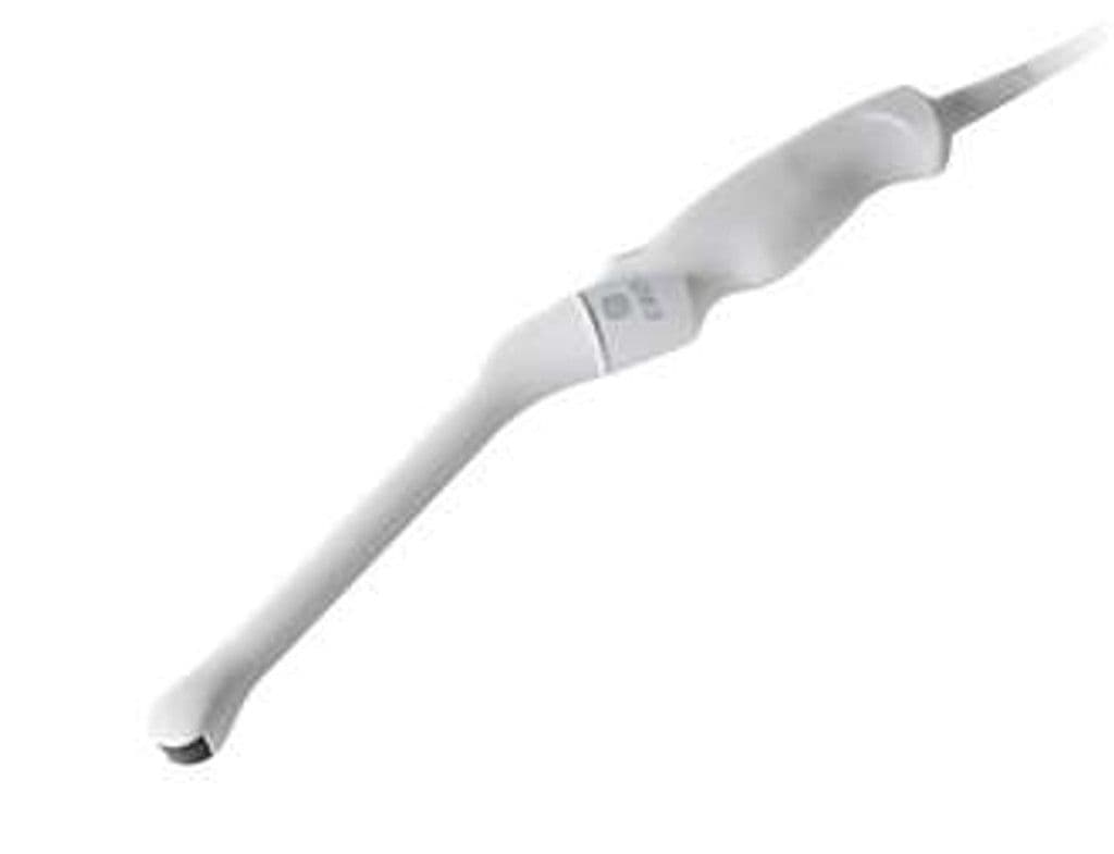
GE E8CS-RS
The E8CS-RS is a micro-convex endocavitary probe with a 4.0 – 10.0 MHz bandwidth and 128-degree field of view built for transvaginal, transrectal, and urological exams. The slim tip eases patient tolerance while the wide aperture captures the uterus, adnexa, prostate, and pelvic floor from standard endocavity approaches. RS-pin compatibility extends its use across GE Versana, LOGIQ, and Vivid portable platforms.

Specifications
Broad endocavitary bandwidth balances near-field resolution for early pregnancy with penetration for deeper pelvic structures.
Wide sector captures bilateral adnexa and full prostate anatomy in a single imaging plane.
Slim profile improves patient comfort during transvaginal and transrectal examinations.
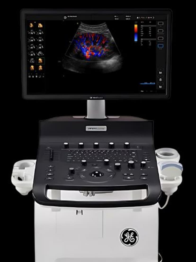
Full Doppler capability for vascular assessment of ovarian, uterine, and prostatic blood flow.
GE RS-Pin connector compatible with Versana, LOGIQ, and Vivid portable systems.
Applications
Transvaginal OB/GYN
The E8CS-RS provides high-resolution transvaginal imaging for early pregnancy dating, follicle monitoring, and endometrial assessment. Its 128-degree field of view captures both adnexa in the same plane, reducing scan time during infertility workups. Color Doppler maps ovarian and uterine artery flow for ectopic pregnancy evaluation and trophoblastic assessment.
Urological Assessment
In urology, the E8CS-RS images the bladder wall, prostate gland, and pelvic floor musculature with the resolution needed for volume calculations and lesion characterization. The 3.0–10.0 MHz range balances near-field prostate detail with deeper bladder penetration, supporting post-void residual measurements and guided biopsy planning.
Transrectal Imaging
For transrectal applications, the probe's compact tip and wide field of view enable full prostate gland visualization including the peripheral zone, transition zone, and seminal vesicles. PW Doppler supports vascularity assessment of suspicious lesions identified during screening exams.
Our Partners


Need this probe for your system?
Request a quote with pricing, compatibility verification, and delivery timeline.