
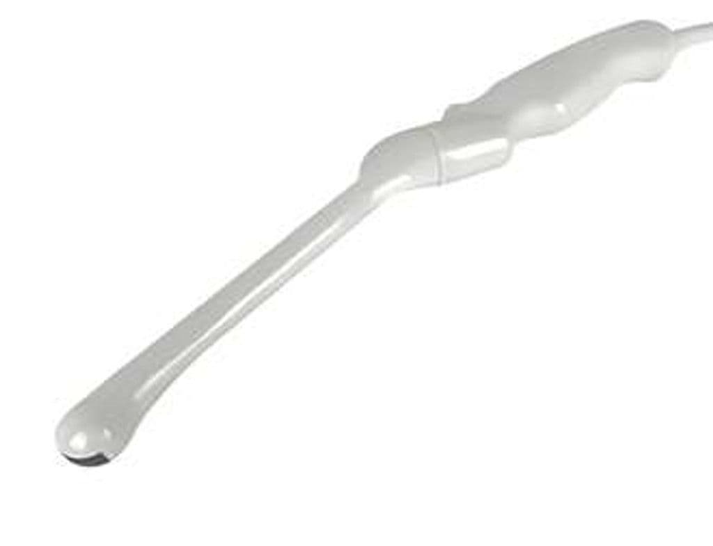
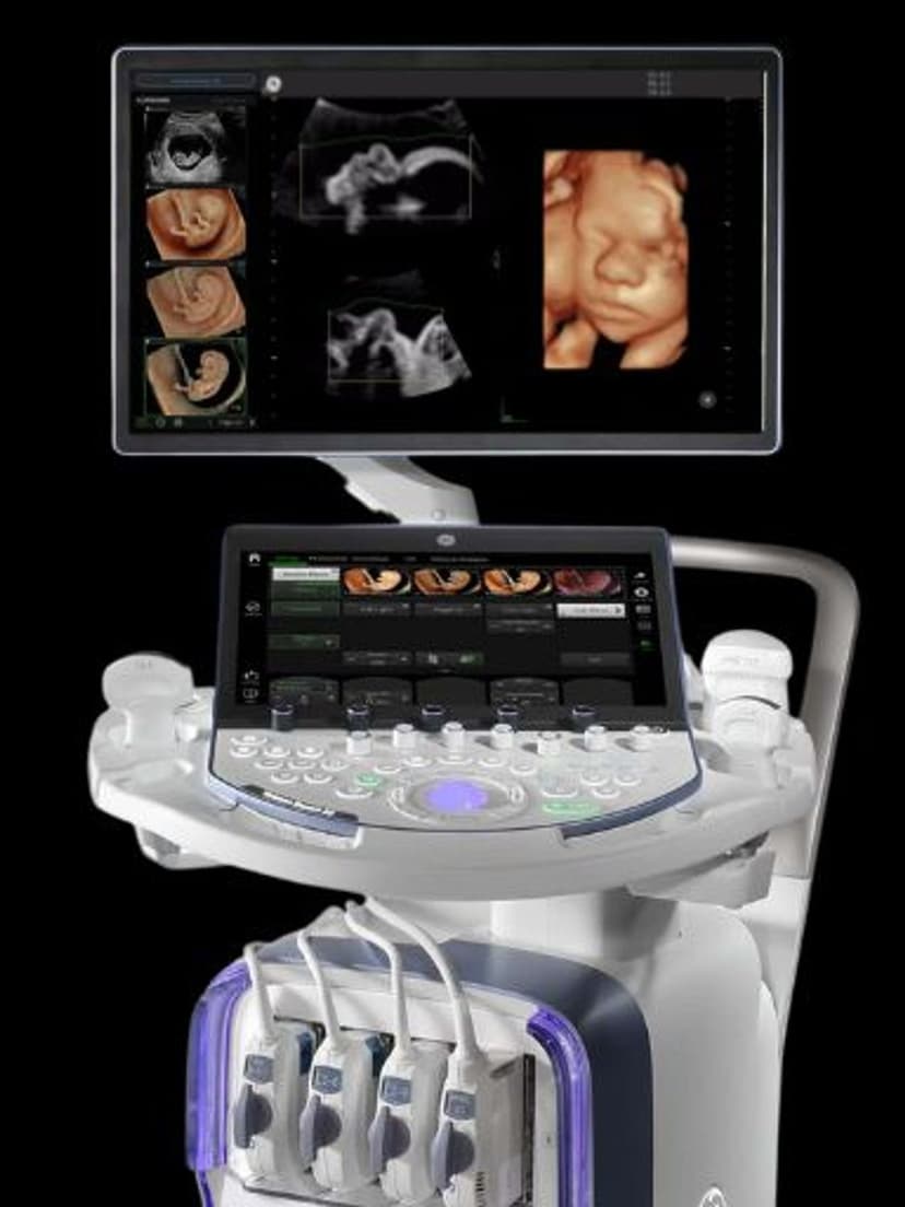
GE IC5-9-D
The IC5-9-D is a micro-convex endocavitary probe with a 3.0 – 9.0 MHz bandwidth and 145-degree field of view designed for GE Vivid and LOGIQ D-Pin flagship platforms. The upper frequency at 9.0 MHz delivers the resolution needed for endometrial detail and early pregnancy structures, while the 3.0 MHz low end maintains penetration for deeper pelvic pathology. The 145-degree sector is wide enough to capture bilateral adnexa without excessive probe manipulation.


Specifications
Extended endocavitary bandwidth resolves fine endometrial and follicular detail while maintaining penetration for deep pelvic structures.
Wide sector angle captures bilateral adnexa and full prostatic anatomy with minimal repositioning.
Full Doppler capability for pelvic vascular assessment and flow quantification.
D-Pin connector for GE Vivid and LOGIQ flagship imaging systems.
Applications
Transvaginal OB/GYN
The IC5-9-D provides transvaginal imaging for early pregnancy dating, adnexal mass characterization, and endometrial assessment. The 9.0 MHz upper frequency resolves endometrial layers, small polyps, and gestational sac detail that lower-frequency endocavitary probes miss. Color Doppler maps corpus luteum vascularity and uterine artery flow indices for preeclampsia screening.
Urological Assessment
For bladder, prostate, and pelvic floor imaging, the IC5-9-D offers the frequency range needed for both superficial urethral detail and deeper bladder wall characterization. Post-void residual measurements and prostate volume calculations use the 145-degree field to capture the entire organ in a standard sagittal or transverse sweep.
Transrectal Imaging
In transrectal applications, the probe images the prostate gland with zonal anatomy detail, supporting lesion localization and guided procedure planning. The wide bandwidth allows operators to optimize frequency for peripheral zone resolution or deeper seminal vesicle visualization depending on the clinical question.
Our Partners


Need this probe for your system?
Request a quote with pricing, compatibility verification, and delivery timeline.