
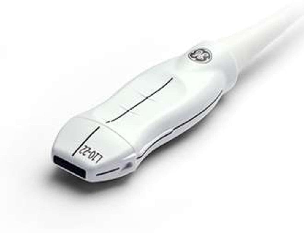
GE L10-22-RS
The L10-22-RS is GE's ultra-high-frequency linear transducer for structures in the first few centimeters of tissue. Its 10.0 – 22.0 MHz range resolves skin layers, tendon fibers, nerve fascicles, and small superficial vessels while the narrow footprint fits fingers, toes, and neonatal extremities. On Venue, LOGIQ P-series, and NextGen LOGIQ e platforms, it supports dermatologic assessment, fine MSK work, and vascular access guidance where standard linears give up near-field detail.

Specifications
Ultra-high frequency reaches sub-100-micron axial resolution for skin layers and superficial tendons.
Full mode set for morphology, flow mapping, and velocity measurement in superficial structures.
Near-field focus maximizes resolution in the first 1–2 cm of tissue depth.
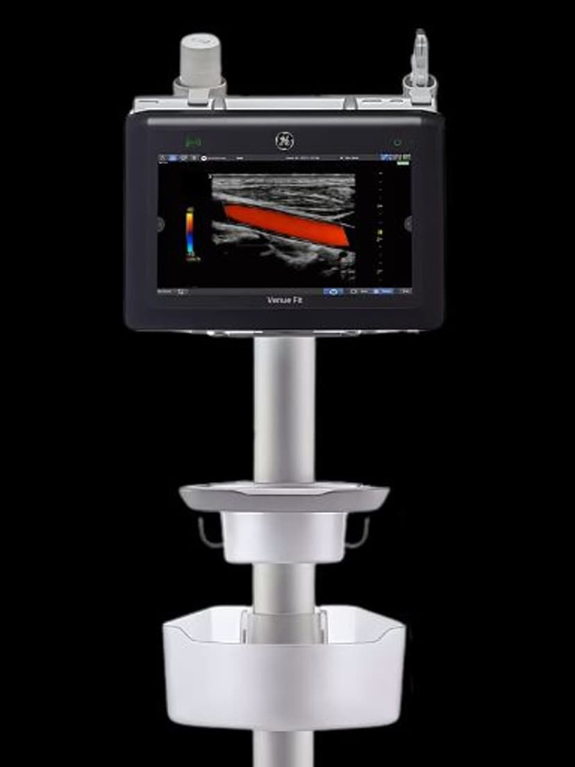
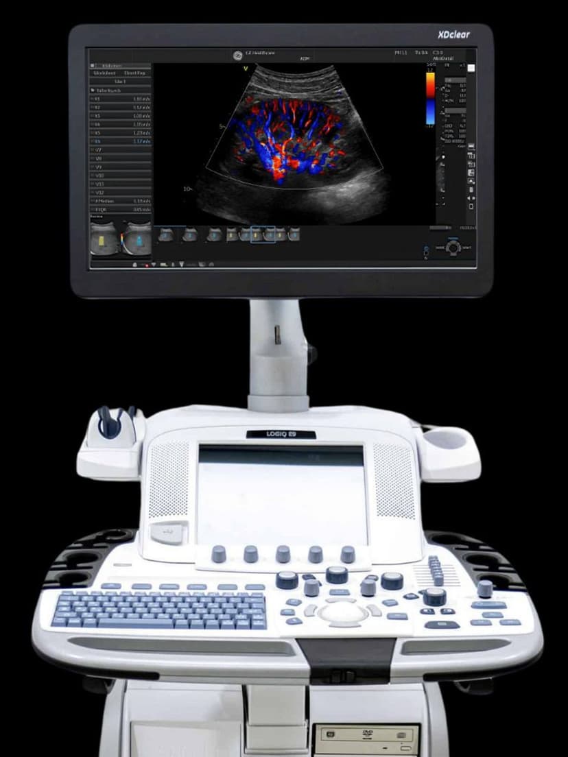
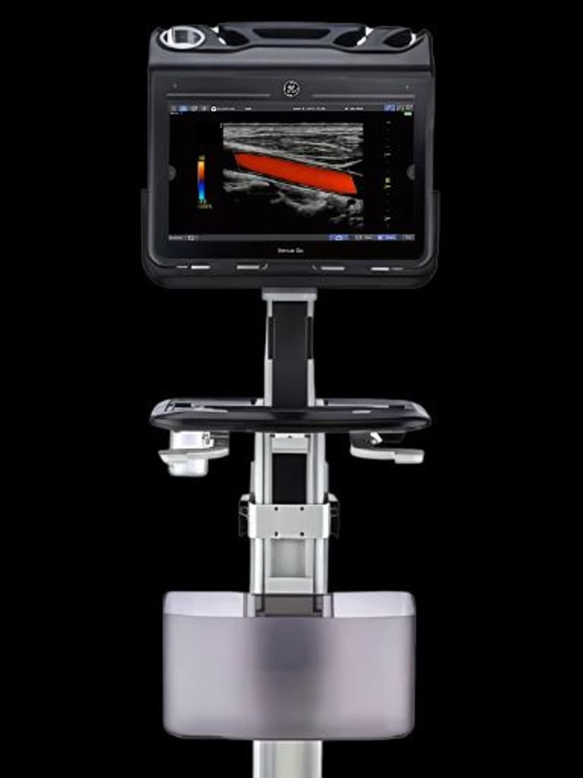
RS-Pin interface connects to GE Venue, LOGIQ P-series, and NextGen LOGIQ e systems.
Applications
Dermatologic Imaging
At 15–20 MHz, the L10-22-RS differentiates epidermis, dermis, and subcutaneous layers for skin lesion depth assessment and procedure planning. Melanoma thickness measurement and margin mapping benefit from the axial resolution at these frequencies. Color Doppler identifies feeder vessels in vascular lesions and inflammatory conditions at the dermal level.
Superficial MSK Assessment
The 7.0–20.0 MHz range resolves individual tendon fibers, nerve fascicles, and joint capsule layers in the hand, wrist, and foot. Trigger finger, de Quervain tenosynovitis, and Morton neuroma assessment benefit from the near-field detail that lower-frequency probes cannot match. The narrow footprint reaches between metacarpals and into the interdigital spaces.
Neonatal & Pediatric Imaging
Superficial structures in neonates, including skin, subcutaneous tissue, and superficial vessels, image with high spatial detail at 12–20 MHz given the minimal tissue depth. Vascular access guidance in premature infants uses the high resolution to identify vessels as small as 1–2 mm. The compact linear footprint fits on small extremities and the neonatal scalp.
Our Partners


Need this probe for your system?
Request a quote with pricing, compatibility verification, and delivery timeline.