
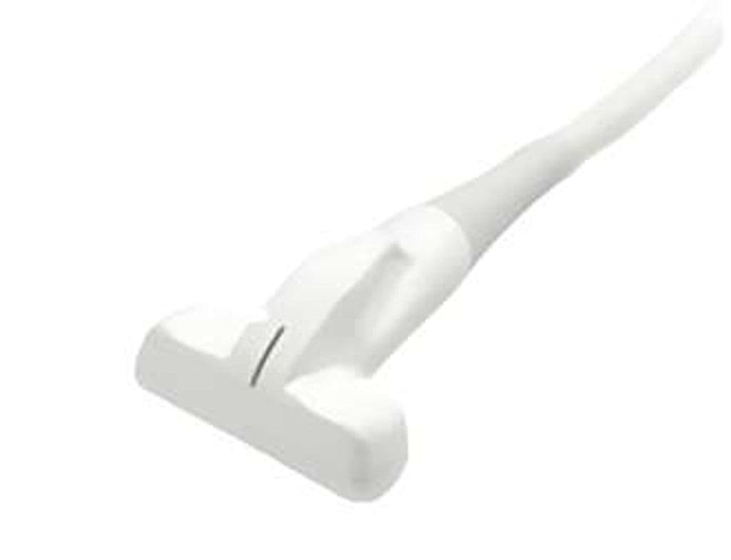
GE L3-9i-RS
The L2.0 – 9.0i-RS is a slim-profile linear transducer purpose-built for intraoperative use. Its 38 mm aperture fits within surgical incisions and between retractors, while the 2.0 – 9.0 MHz bandwidth images structures from the skin surface down to 10 cm depth. Surgeons use it for real-time guidance during vascular repair, tumor localization, and intraoperative Doppler assessment of graft patency and organ perfusion.
Compatible Systems

Specifications
Bandwidth spans superficial detail at 9 MHz to 10 cm penetration at 3 MHz for versatile intraoperative use.
Compact aperture fits within surgical incisions and between retractors during open procedures.
Reaches mid-depth structures encountered during abdominal and vascular surgical procedures.
Full Doppler capability supports intraoperative graft patency checks and flow assessment.
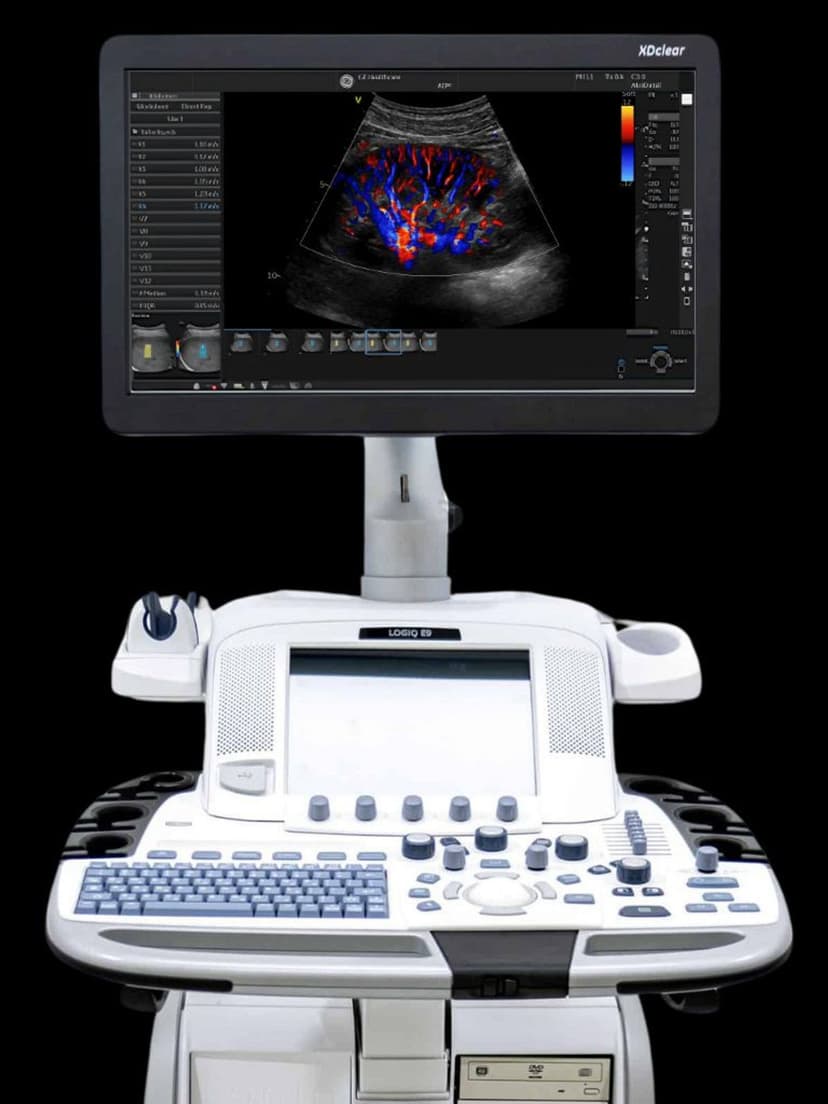
RS-Pin connector compatible with GE LOGIQ-series imaging systems.
Applications
Intraoperative Imaging
The 38 mm aperture and slim housing fit directly into the surgical field for real-time tissue assessment. Surgeons verify tumor margins, locate structures relative to planned resection lines, and confirm complete excision before wound closure. The 3–9 MHz range covers both superficial skin-level structures and deeper organ parenchyma encountered during open abdominal and thoracic procedures.
Vascular Assessment
Intraoperative Doppler confirms graft patency after bypass and validates flow restoration during carotid endarterectomy. Color and PW Doppler modes detect residual stenosis or dissection flaps that may require immediate revision. The probe also supports peripheral vascular mapping in outpatient settings when used on LOGIQ systems.
Musculoskeletal & Soft Tissue Imaging
Lower-frequency operation at 3–5 MHz provides deeper penetration than standard high-frequency linear probes, reaching the hip joint, deep gluteal space, and thigh compartments. Higher frequencies within the 9 MHz range resolve superficial tendons and ligaments. This dual capability suits orthopedic practices that image both deep joints and superficial structures.
Our Partners


Need this probe for your system?
Request a quote with pricing, compatibility verification, and delivery timeline.