
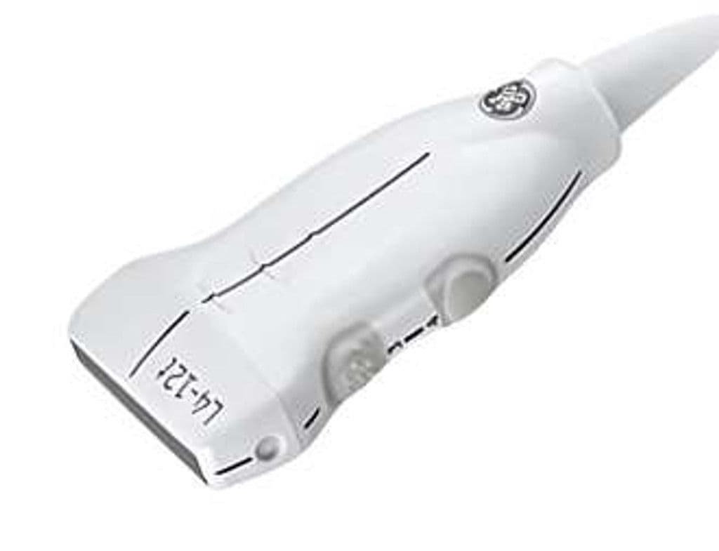
GE L4-12t-RS
The L4-12t-RS covers superficial to mid-depth anatomy across a 4.2 – 13.0 MHz bandwidth, reaching structures up to 6 cm deep through a 12.7 x 47.1 mm footprint. Configurable thumb buttons let operators switch presets and freeze frames without reaching for the console. Compatible with Venue, LOGIQ, and NextGen LOGIQ e platforms via the RS-Pin connector.

Specifications
Broad bandwidth covers pediatric through adult superficial imaging without switching probes.
Wide aperture captures thyroid lobes and breast regions in fewer image sweeps.
Optimized for superficial and mid-depth structures in small parts and MSK exams.
Full diagnostic mode set for tissue morphology and vascular hemodynamics.
Programmable thumb buttons allow one-handed preset changes during active scanning.
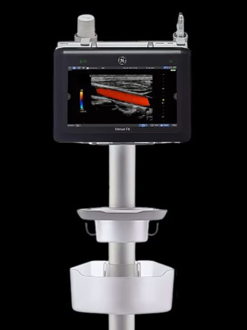
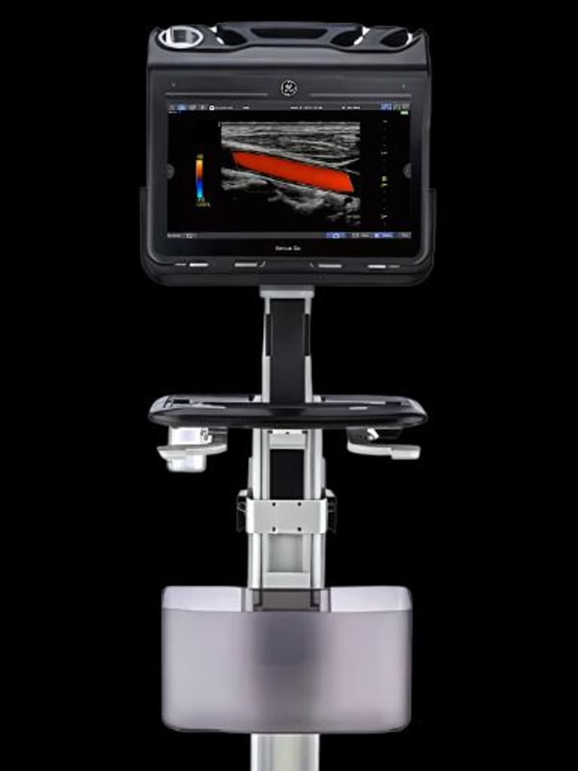
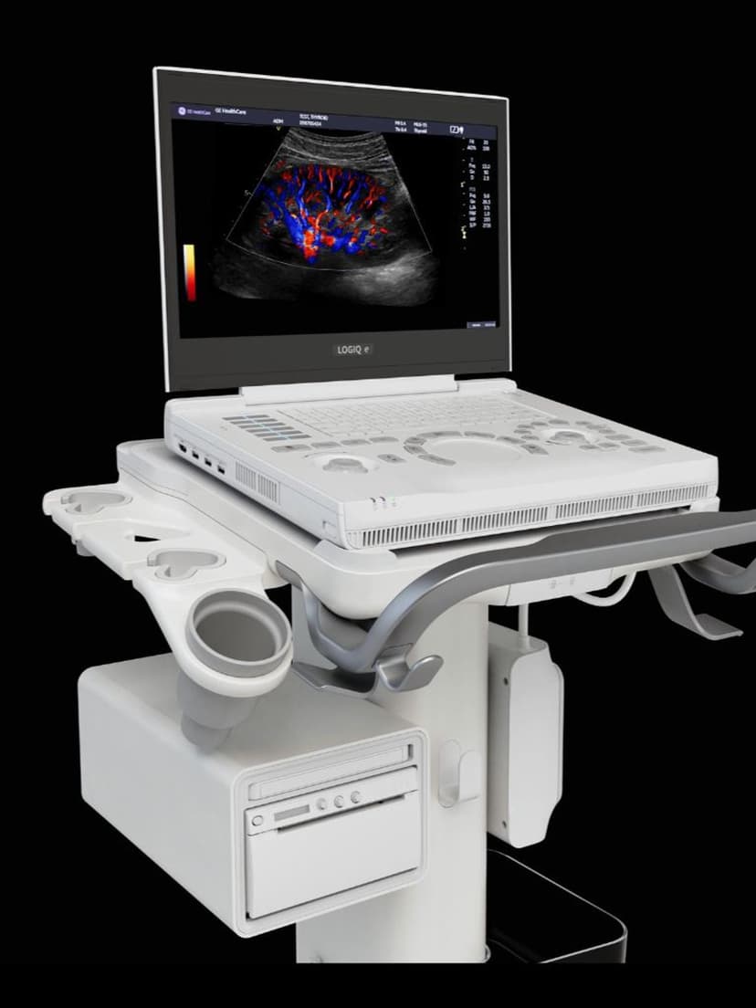
RS-Pin interface connects to GE Venue, LOGIQ P-series, and NextGen LOGIQ e systems.
Applications
Breast & Thyroid Imaging
The 12 MHz upper range resolves sub-centimeter thyroid nodules and breast lesions for TI-RADS and BI-RADS classification. The 47.1 mm aperture captures most thyroid lobes without panoramic stitching, and the slim footprint seats firmly in the supraclavicular fossa and intercostal spaces. Color Doppler maps vascularity patterns used in nodule risk stratification.
Musculoskeletal Assessment
High-frequency operation at 8–12 MHz differentiates tendon fiber bundles, ligament borders, and joint effusions in shoulder, elbow, wrist, and ankle exams. The 4.2 MHz low end extends reach to deeper structures like the supraspinatus in larger patients. Dynamic scanning during movement reveals impingement and subluxation in real time.
Pediatric Imaging
The compact linear footprint and broad bandwidth suit neonatal cranial, abdominal, and hip imaging where both resolution and penetration matter. Configurable buttons reduce scanning time in uncooperative patients by keeping key functions at the operator's fingertips. PW Doppler supports vascular assessment in pediatric renal and hepatic studies.
Our Partners


Need this probe for your system?
Request a quote with pricing, compatibility verification, and delivery timeline.