
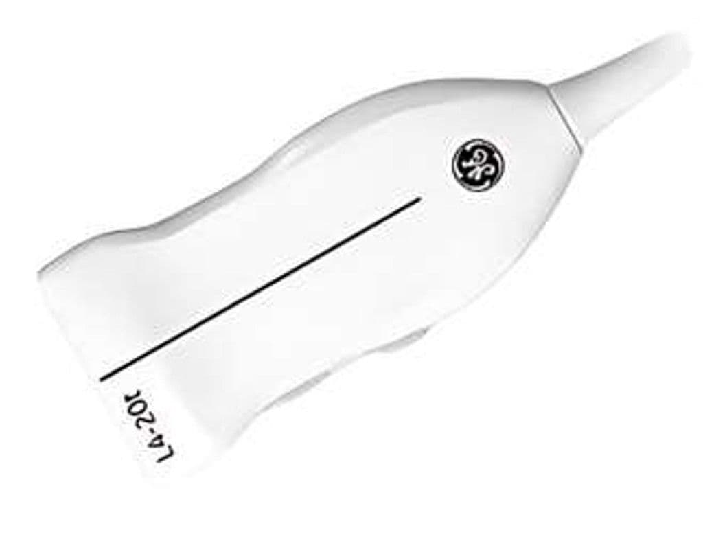
GE L4-20t-RS
The L4-20t-RS pairs XDclear crystal technology with an ultra-wideband 5.0 – 20.0 MHz bandwidth, delivering sub-millimeter resolution on superficial tendons and nerves while retaining penetration for deeper vascular structures. Center-line markers on the probe face support needle-guided procedures, and four configurable buttons give operators single-press access to frequently used functions without returning to the console.
Specifications
Ultra-wideband range covers superficial MSK detail through mid-depth vascular imaging in a single transducer.
GE's single-crystal technology improves sensitivity and bandwidth over conventional piezoelectric elements.
Full 2D and Doppler suite supports diagnostic and interventional workflows.
Programmable buttons reduce console interaction during sterile or time-sensitive procedures.
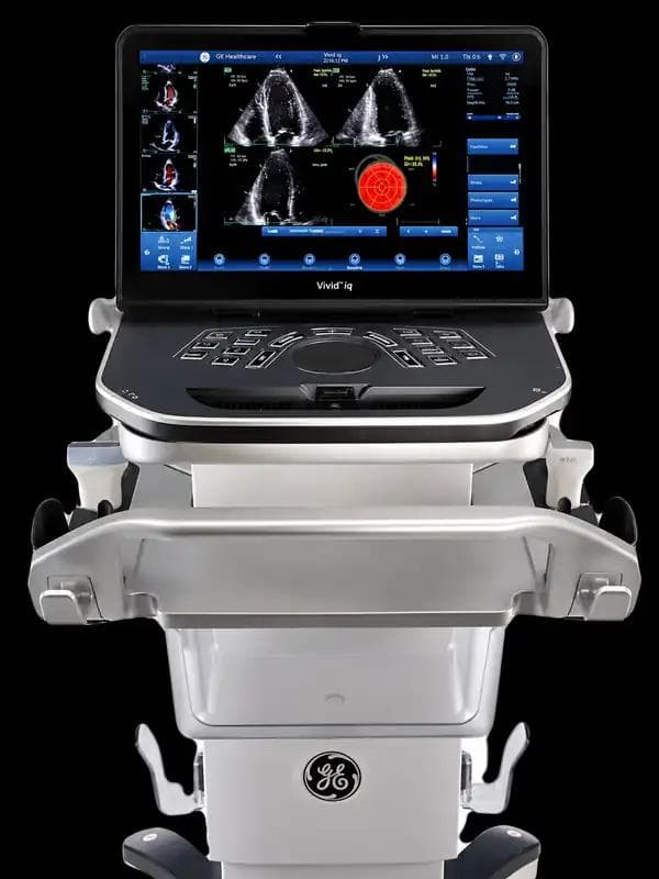
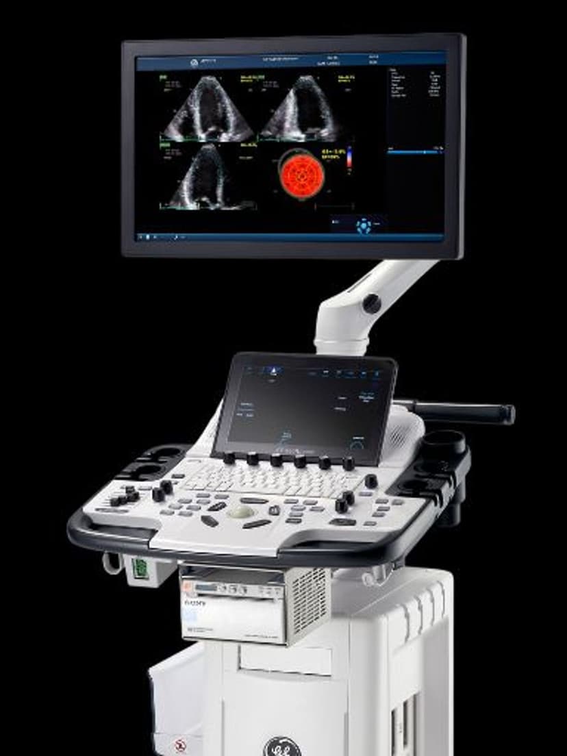
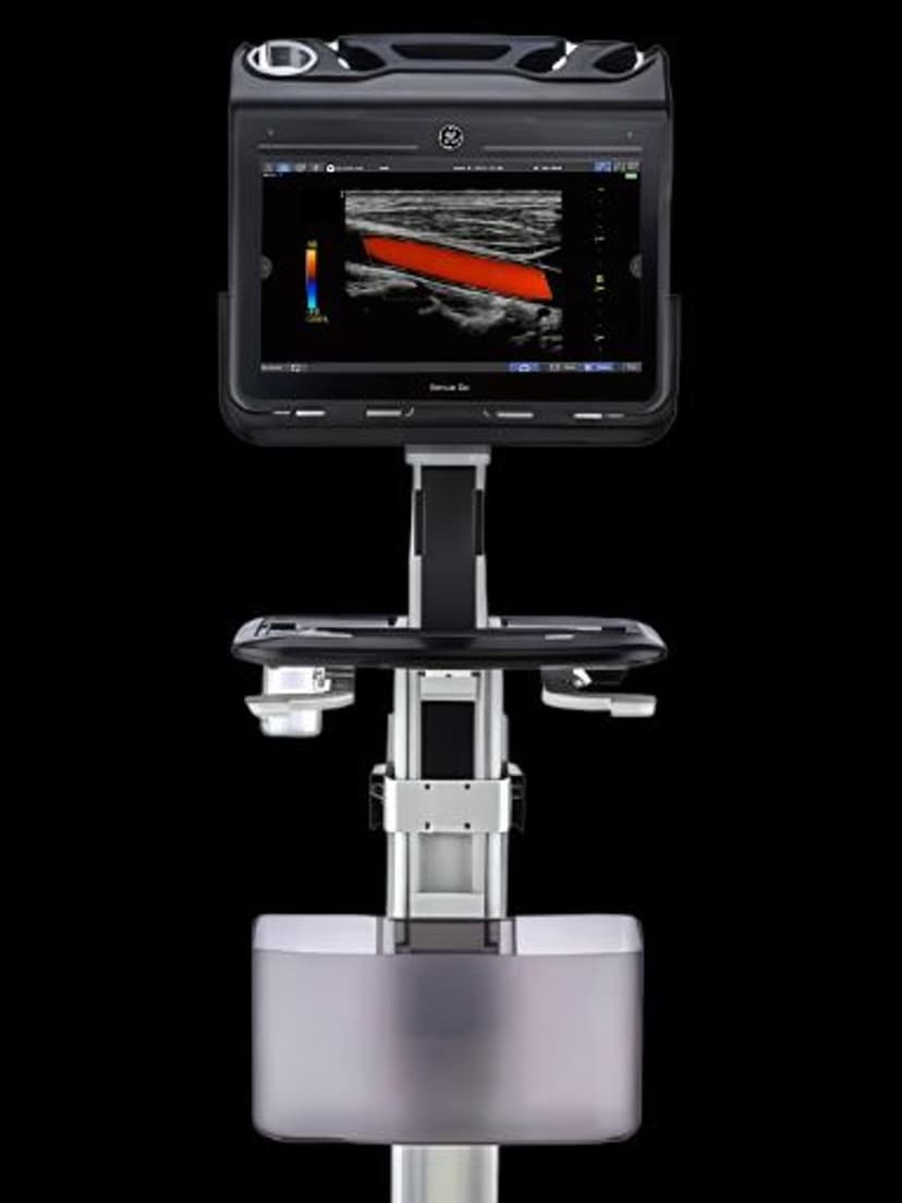
RS-Pin connector is compatible with GE systems that use the standard RS-series interface.
Applications
Musculoskeletal Imaging
Resolves fine tendon fiber architecture, ligament tears, and joint effusions at frequencies up to 20 MHz. The wide bandwidth allows operators to shift between high-resolution superficial imaging and deeper muscle or joint assessments without switching probes. B-mode and Color Doppler support detection of hyperemia around inflamed tendons and bursae.
Ultrasound-Guided Nerve Blocks
Center-line markers on the probe face align the needle path with the ultrasound plane for in-plane approaches. High-frequency imaging differentiates nerve fascicles from surrounding fascia, supporting brachial plexus, femoral, and sciatic blocks. The slim footprint fits confined spaces around the neck and axilla.
Peripheral Vascular Assessment
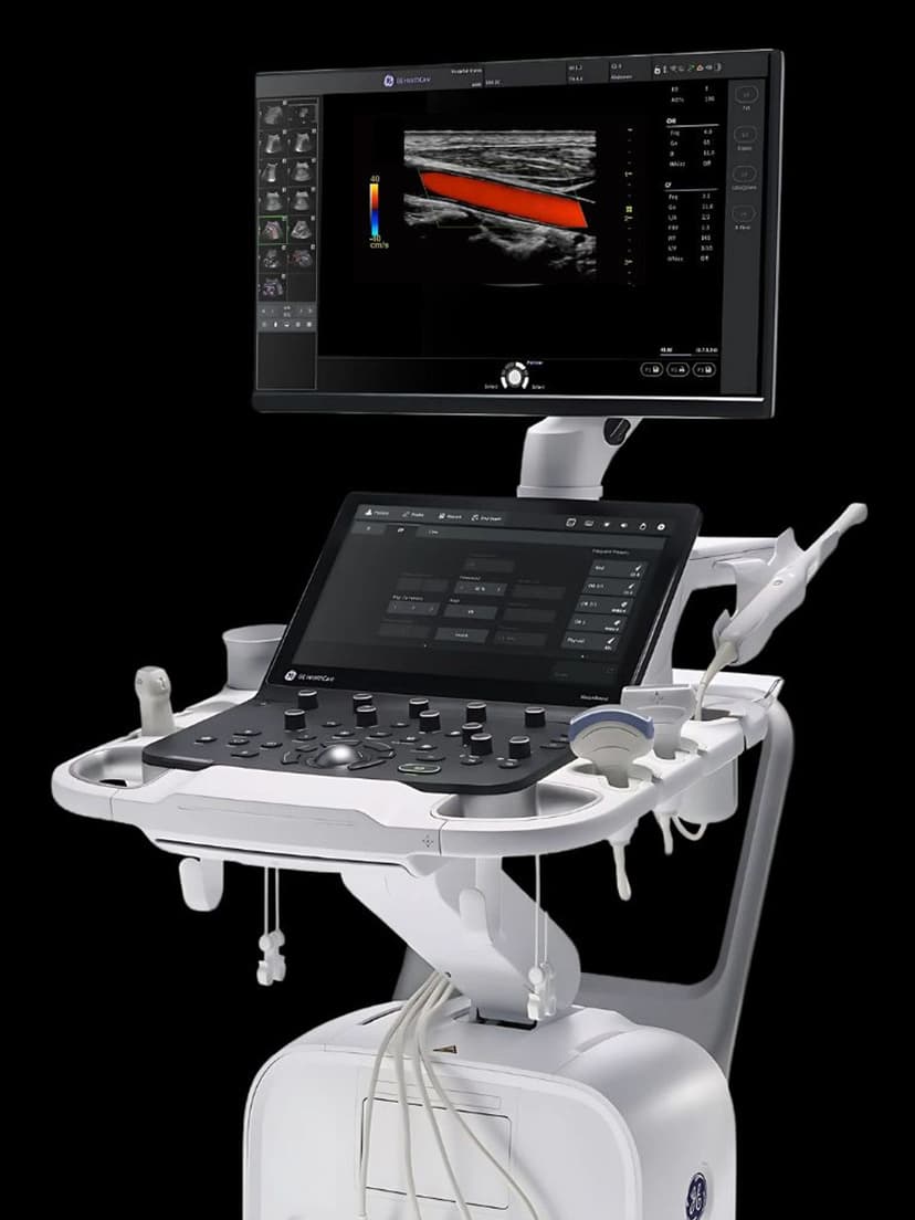
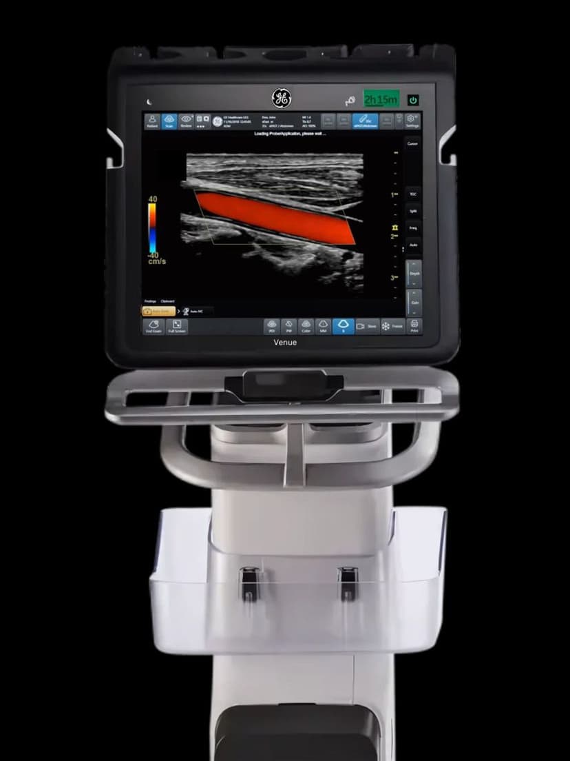
Color and PW Doppler map flow in carotid, radial, and pedal arteries with the resolution needed to grade stenosis and detect thrombus. The broad bandwidth accommodates both superficial vessels and mid-depth structures such as the popliteal artery without transducer changes.
Our Partners


Need this probe for your system?
Request a quote with pricing, compatibility verification, and delivery timeline.