
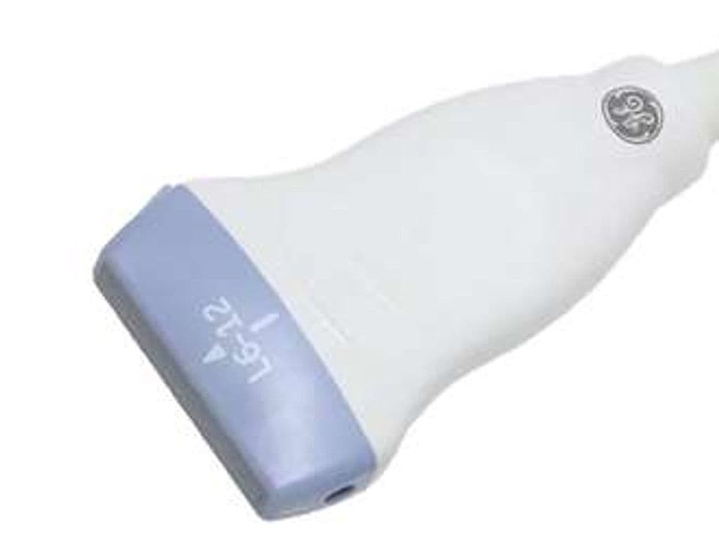
GE L6-12-RS
The L6-12-RS provides a 4.0 – 13.0 MHz bandwidth through a 38.4 mm aperture, balancing penetration depth (up to 6 cm) with the resolution needed for superficial structure assessment. The slim linear footprint accesses tight anatomical regions including the supraclavicular fossa and posterior knee. Compatible with eight RS-Pin systems spanning the Versana, LOGIQ, and Vivid T9 lines, making it one of GE's most widely deployed linear transducers.

Specifications
Broad bandwidth covers vascular depth studies through superficial small parts imaging in one probe.
Moderate aperture fits thyroid lobes and vessel segments while maintaining a compact footprint.
Covers superficial and mid-depth structures typical of MSK, small parts, and vascular exams.
Standard diagnostic mode set for morphology, flow mapping, and velocity quantification.
RS-Pin interface connects to GE Versana, LOGIQ P-series, and Vivid T9 systems.
Applications
Vascular Imaging
The 4 MHz low end reaches deeper vessels like the common femoral and subclavian arteries, while the 12 MHz upper range resolves superficial structures including the carotid intima-media complex. The 38.4 mm aperture provides adequate vessel length for spectral Doppler sampling with proper angle correction. Color and PW Doppler support velocity measurement and stenosis grading across the peripheral vascular tree.
Small Parts & Thyroid Imaging
High-frequency operation between 8–12 MHz differentiates thyroid nodule characteristics including echogenicity, margins, and calcifications for TI-RADS scoring. The 38.4 mm field covers most thyroid lobes in a single longitudinal sweep. Breast lesion assessment benefits from the same frequency range for BI-RADS classification and biopsy guidance.
Musculoskeletal Assessment
Dynamic scanning of tendons, ligaments, and joints at the shoulder, elbow, wrist, and ankle uses the 8–12 MHz range for fiber-level resolution. The slim profile slides between bony landmarks at the ankle and wrist where bulkier probes cannot maintain surface contact. Color Doppler detects hyperemia in tendinopathy, bursitis, and post-operative monitoring.
Our Partners


Need this probe for your system?
Request a quote with pricing, compatibility verification, and delivery timeline.