
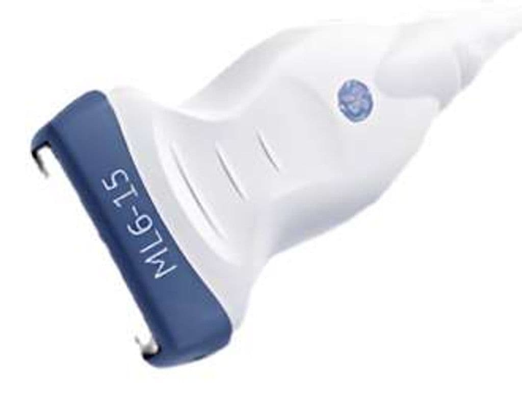
GE ML6-15-D
The ML6-15-D uses matrix array architecture to electronically focus in both the elevation and lateral planes, producing uniformly sharp images across the full 49.6 mm field of view. The 4.0 – 15.0 MHz bandwidth covers everything from deep vascular structures to sub-millimeter thyroid nodules without switching probes. On LOGIQ Fortis, LOGIQ E10, and Vivid E95 systems, the matrix element layout supports coded excitation and spatial compounding for improved tissue differentiation.


Specifications
Wideband range images deep vascular structures through superficial thyroid and breast tissue in one probe.
Matrix architecture enables electronic focusing in both elevation and lateral planes for uniform image quality.
Wide aperture captures full thyroid lobes and vascular segments in a single view.
Optimized for superficial and mid-depth structures commonly imaged with high-frequency linear probes.
Standard diagnostic mode set for vascular hemodynamics and tissue morphology.
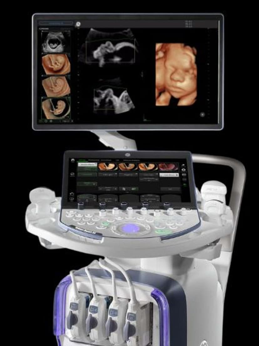
D-Pin interface connects to GE's flagship LOGIQ and Vivid systems.
Applications
Vascular Imaging
Matrix focusing sharpens carotid plaque borders and vessel wall detail for accurate intima-media thickness measurement. The 4.5 MHz low end reaches the subclavian and deeper femoral vessels, while 15 MHz resolves superficial temporal and radial arteries. PW Doppler angle correction and spectral analysis support hemodynamic quantification across the full vascular tree.
Breast & Thyroid Imaging
High-frequency operation at 10–15 MHz resolves thyroid nodules and breast lesions for TI-RADS and BI-RADS scoring. The 49.6 mm aperture captures the full thyroid lobe in a single image, reducing the need for panoramic stitching. Elastography compatibility on supported systems adds stiffness data for lesion characterization.
Musculoskeletal Imaging
Matrix beam focusing maintains lateral resolution across the image, which improves visualization of tendon fiber continuity and ligament integrity compared to single-row linear arrays. Dynamic scanning at the shoulder, elbow, and ankle benefits from the wide aperture and consistent image quality. Color Doppler detects hyperemia in tendinopathy and post-surgical sites.
Our Partners


Need this probe for your system?
Request a quote with pricing, compatibility verification, and delivery timeline.