
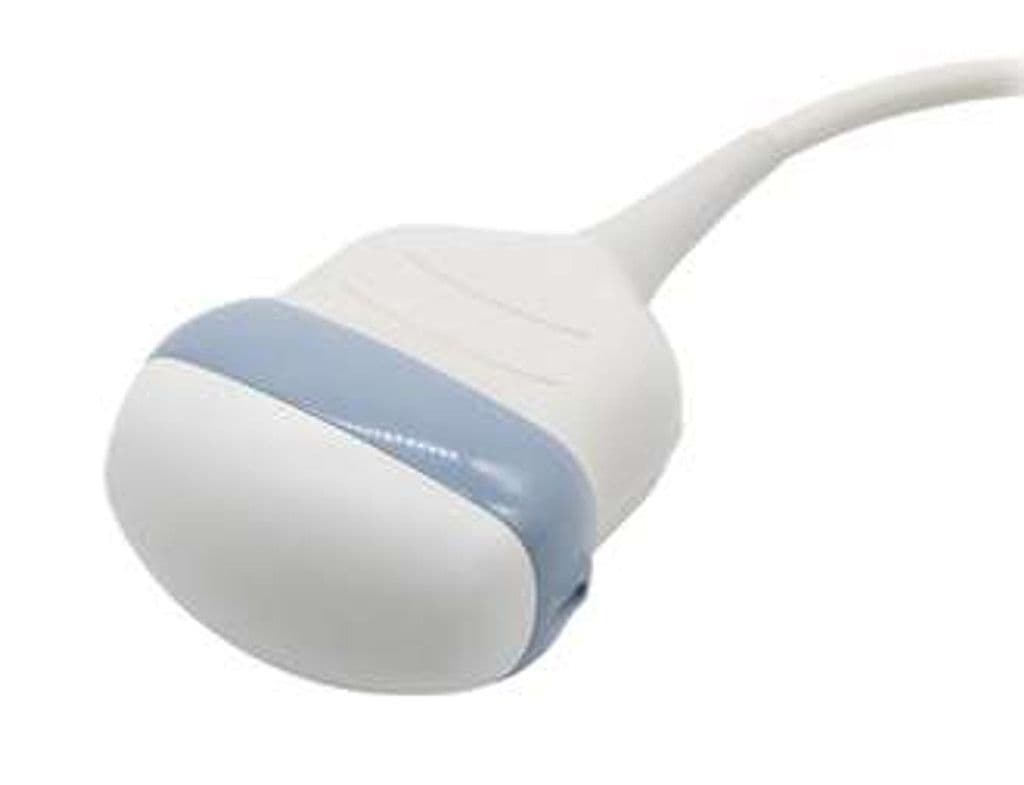
GE RAB6-RS
The RAB6-RS is GE's primary 4D volume convex probe for the Voluson women's health platform, covering 2.0–8.0 MHz with a 90-degree B-mode and 90x85-degree volume field of view. It handles the full spectrum of obstetric imaging from first-trimester screening through third-trimester growth scans, with HDlive-compatible volume acquisition for surface-rendered fetal imaging. The ultralight construction reduces operator fatigue during high-volume OB scan days.
Compatible Systems
Specifications
Wide bandwidth supports obstetric imaging from high-BMI patients to detailed fetal anatomy in thinner patients.
Standard convex sector covers abdominal and obstetric anatomy in a single sweep.
Large volume sector captures complete fetal anatomy for HDlive rendering and STIC cardiac evaluation.
Full Doppler and 4D volume modes support both structural and hemodynamic fetal assessment.
RS-Pin interface for the Voluson women's health platform.
Applications
Obstetric 3D/4D Imaging
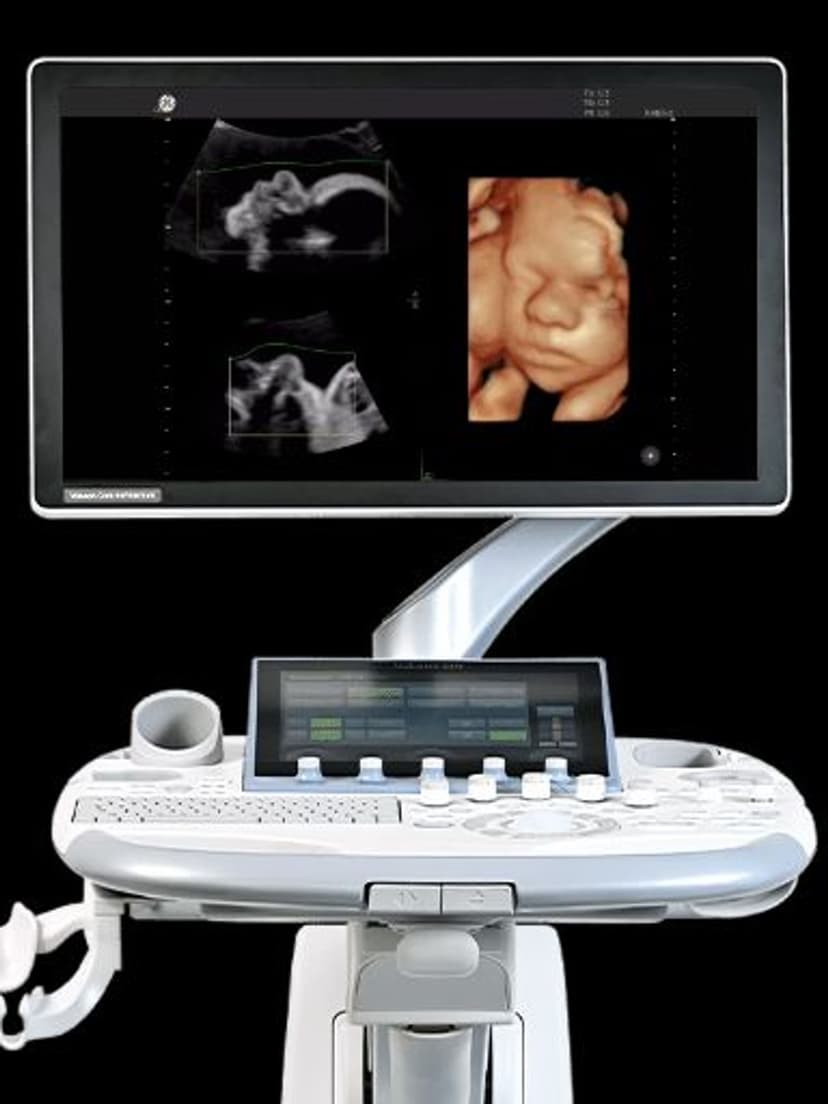
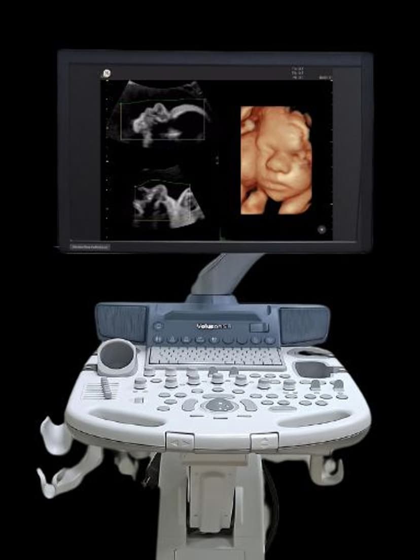
The RAB6-RS is the primary volume acquisition probe for Voluson obstetric workflows. It captures the datasets used for HDlive surface rendering, STIC cardiac volumes, and tomographic fetal brain imaging. The 2.0 MHz low end penetrates through increased maternal BMI, while the 8.0 MHz high end resolves fetal facial features, digits, and spine detail in thinner patients. Real-time 4D renders fetal movement and facial expressions for parent bonding and behavioral assessment.
Abdominal Imaging
Beyond obstetrics, the RAB6-RS functions as a general-purpose abdominal probe with volume capability. The curved array geometry and 90-degree field of view cover liver, gallbladder, kidneys, and spleen in standard scanning planes. Volume acquisition adds multiplanar reconstruction for complex lesion characterization and 3D organ volume measurement.
Gynecologic Assessment
For transabdominal GYN imaging, the probe provides uterine and ovarian visualization with 3D volume capability for fibroid mapping and endometrial assessment. The 2.0–8.0 MHz bandwidth accommodates both deeper pelvic imaging through a full bladder and higher-resolution views of anterior structures. Volume datasets support coronal uterine views for Mullerian anomaly classification.
Our Partners


Need this probe for your system?
Request a quote with pricing, compatibility verification, and delivery timeline.