
GE RIC5-9-D
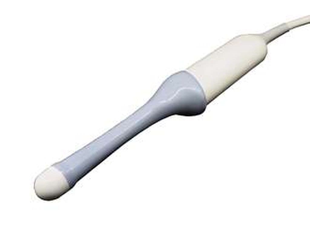
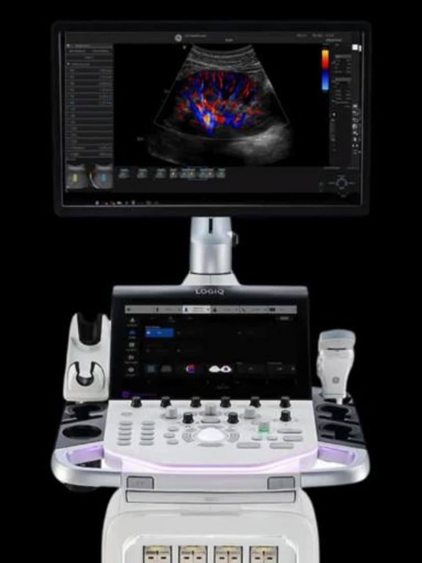
The RIC5-9-D brings real-time 4D volume imaging to endocavitary exams, covering 4.0–9.0 MHz with a 179-degree B-mode field of view. On the LOGIQ E10, Totus, and Fortis platforms, it delivers volumetric rendering of uterine anatomy, follicular structures, and IUD placement confirmation that 2D endocavitary probes cannot provide. The micro-convex element geometry preserves the slim profile needed for patient comfort while adding the sweep mechanism required for automated volume acquisition.


Specifications
Wideband endocavitary frequency range covers near-field reproductive structures through deeper pelvic anatomy.
Near-panoramic field captures full uterine and prostatic anatomy with minimal repositioning.
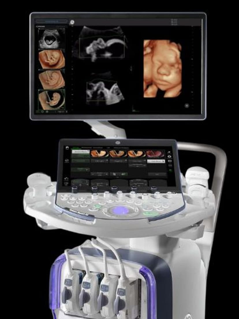
4D volume acquisition adds coronal plane reconstruction and tomographic imaging to standard 2D endocavitary workflows.
D-Pin connector for LOGIQ premium imaging platforms.
Applications
3D/4D Transvaginal OB/GYN
The RIC5-9-D enables volume-rendered transvaginal imaging for coronal uterine plane visualization, a view impossible with conventional 2D endocavitary probes. This aids Mullerian anomaly classification, IUD localization, and detailed assessment of endometrial polyps. Real-time 4D acquisition captures uterine peristalsis and provides tomographic ultrasound imaging (TUI) slices for systematic evaluation of complex pathology.
Urologic Imaging
In transrectal applications, the 4D capability supports volumetric prostate assessment including gland volume measurement and targeted biopsy planning. The 179-degree field of view captures the entire gland in a single volume dataset, enabling multiplanar reconstruction for lesion localization relative to the capsule, urethra, and neurovascular bundles.
Obstetric Assessment
For early pregnancy, the RIC5-9-D provides volumetric imaging of embryonic and early fetal anatomy when transabdominal access is limited. The 4.0–9.0 MHz range resolves nuchal translucency and early anatomic structures with the near-field detail required during the first trimester. Volume datasets allow offline review and second-opinion consultation.
Our Partners


Need this probe for your system?
Request a quote with pricing, compatibility verification, and delivery timeline.