
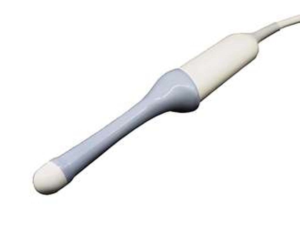
GE RIC5-9A-RS
The RIC5-9A-RS adds real-time 4D volume acquisition to an endocavity microconvex design, enabling 3D/4D transvaginal imaging of the uterus, ovaries, and early fetal anatomy. The 146-degree B-mode field of view and 146 x 84-degree volume scan angle provide wide pelvic coverage from the intracavitary position. The 20.96 x 23.35 mm footprint balances patient comfort against the element density needed for volume rendering, and the 4.2–10 MHz bandwidth supports both deep pelvic mass assessment and high-resolution follicular monitoring.

Specifications
Wideband range covers deep pelvic masses at the low end and high-resolution follicular imaging at the upper range.
Extra-wide sector captures the full uterine fundus and bilateral ovaries in one view.
Volume sweep angle produces 3D/4D datasets wide enough for full uterine and ovarian coverage.
Compact tip balances patient comfort against the element density needed for volume imaging.
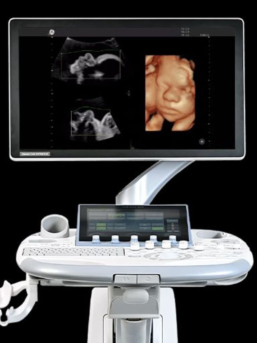
Real-time 4D mode enables HDlive rendering and automated follicle counting on compatible systems.
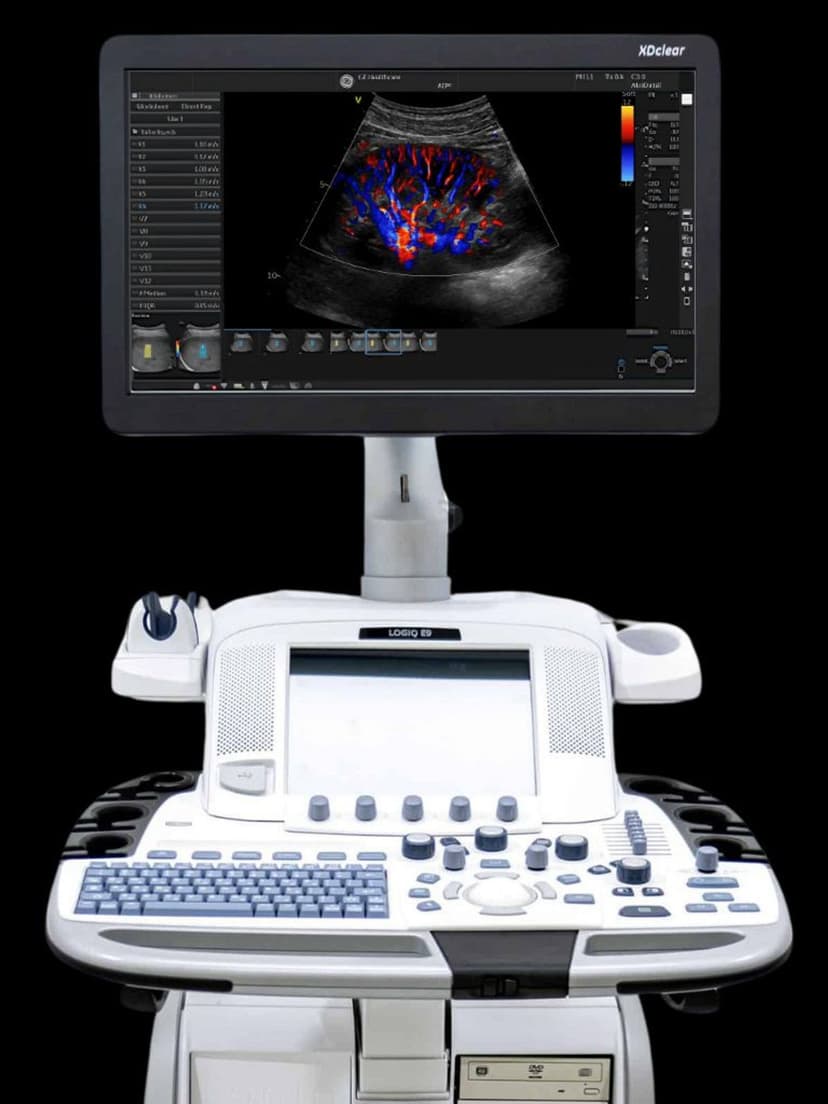
RS-Pin interface connects to Voluson, LOGIQ, and Versana platforms for 3D/4D endocavity imaging.
Applications
Transvaginal 3D/4D OB/GYN Imaging
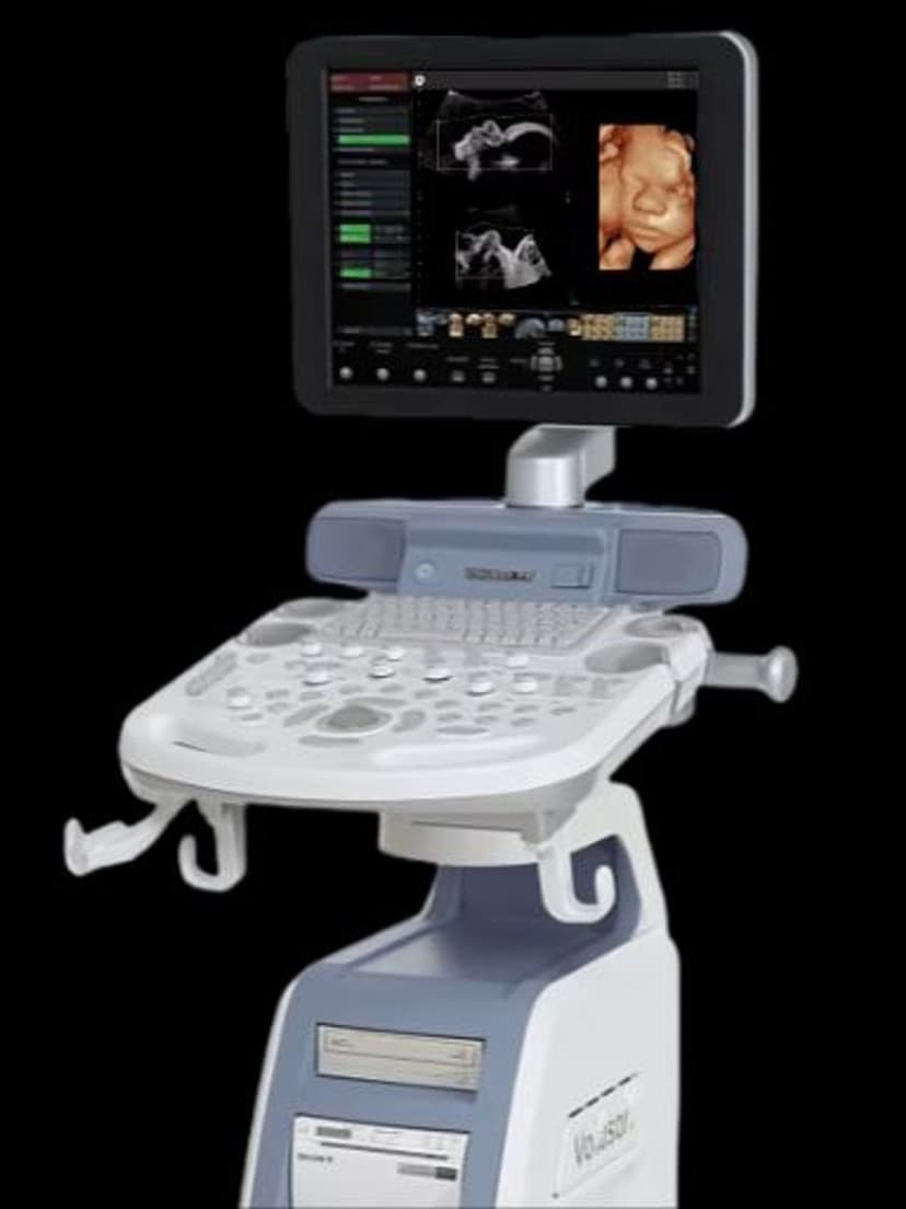
Volume acquisition generates coronal uterine views that identify septate, bicornuate, and arcuate anomalies without the need for MRI. The 146-degree field captures the full uterine fundus and both ovaries in a single sweep. HDlive-compatible datasets on Voluson systems produce 3D surface rendering of early fetal anatomy, including face and spine, from the first trimester.
Fertility & Follicular Monitoring
At 8–10 MHz, the RIC5-9A-RS resolves individual follicles for count and diameter measurement during IVF stimulation cycles. Automated follicle count tools on compatible systems reduce inter-observer variability and speed up the monitoring exam. 3D volume mode stores the full ovarian dataset for offline review, preventing recall visits when borderline follicle counts need re-evaluation.
Pelvic Floor & Transrectal Assessment
The wide sector angle captures levator ani morphology and pelvic organ position during dynamic maneuvers. Volume rendering reconstructs the hiatal area in the axial plane for prolapse grading and surgical planning. Transrectal positioning images the prostate and seminal vesicles with the same probe, and the 4.2 MHz low end reaches deeper pelvic structures through either approach.
Our Partners


Need this probe for your system?
Request a quote with pricing, compatibility verification, and delivery timeline.