
GE Ultrasound Feature
LVO Contrast
ContrastLVO Contrast (Left Ventricular Opacification) uses injected ultrasound contrast agents to enhance the echogenicity of blood within the left ventricle, creating a clear boundary between the blood pool and the ventricular wall. This is important for patients with poor acoustic windows where standard B-mode imaging cannot adequately define endocardial borders, such as patients with obesity, COPD, or post-surgical anatomy. With opacified blood clearly outlining the ventricle, clinicians can accurately assess wall motion, measure ejection fraction, and detect intracardiac thrombi that would otherwise be missed. LVO Contrast imaging is used in echocardiography labs and cardiology practices where a significant percentage of studies produce suboptimal images without contrast enhancement.

Key Benefits
Why LVO Contrast matters
Definitive endocardial border visualization
The contrast-enhanced blood pool creates a sharp boundary against the myocardium, making endocardial borders visible in patients where standard imaging fails. This converts technically inadequate studies into diagnostic ones, reducing the need for repeat exams or alternative imaging.
Accurate ejection fraction in difficult-to-image patients
Patients with obesity, COPD, or chest wall deformities often produce echocardiograms where EF measurement is unreliable due to poor border definition. LVO Contrast provides the border clarity needed for accurate biplane Simpson's EF calculation in these patients.
Improved detection of left ventricular thrombi
Small or mural thrombi in the left ventricle are commonly missed on standard echocardiography. Contrast fills the ventricular cavity and makes thrombi visible as filling defects, catching a finding that directly changes anticoagulation management.
Fewer non-diagnostic studies and downstream imaging
When an echocardiogram is non-diagnostic due to poor image quality, the patient is often sent for cardiac MRI or a repeat study. LVO Contrast rescues many of these studies at the time of the initial exam, avoiding the cost, scheduling burden, and patient inconvenience of additional imaging.
About LVO Contrast
Left ventricular opacification works by intravenously injecting a microbubble-based contrast agent that enhances the ultrasound signal from blood. The microbubbles resonate at diagnostic ultrasound frequencies and produce a strong backscatter signal, making the blood pool within the left ventricle appear bright on the display. This creates a sharp contrast between the opacified blood and the myocardium, allowing clear visualization of endocardial borders that may be invisible on unenhanced imaging. The GE Vivid system's LVO Contrast mode optimizes transmit power, gain, and processing algorithms specifically for contrast-enhanced imaging to maintain bubble integrity and maximize border definition. Clinical guidelines from the American Society of Echocardiography recommend the use of contrast agents when two or more contiguous endocardial segments cannot be visualized on standard imaging. In practice, studies suggest that 10-20% of echocardiograms have suboptimal image quality that warrants contrast enhancement. LVO Contrast also improves the detection of left ventricular thrombi, where contrast fills the ventricle and makes thrombi visible as filling defects. Without contrast, small or mural thrombi are frequently missed on standard echocardiography.
Availability
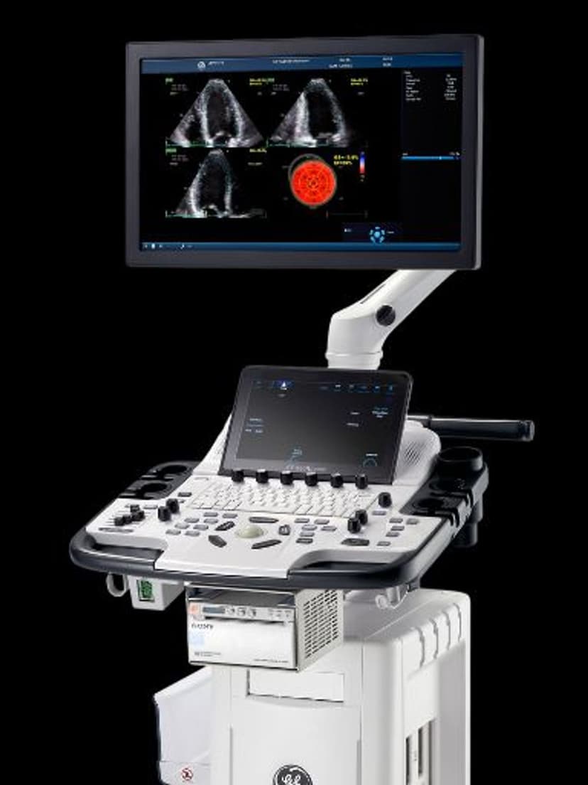
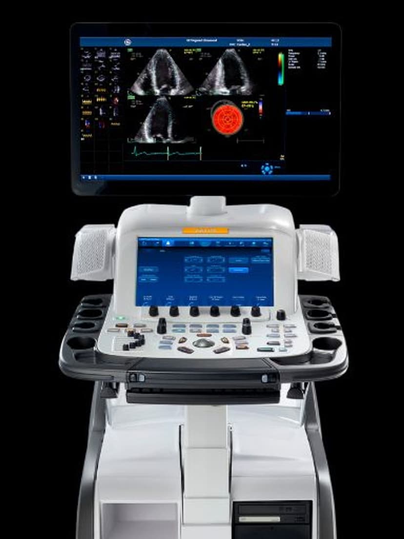
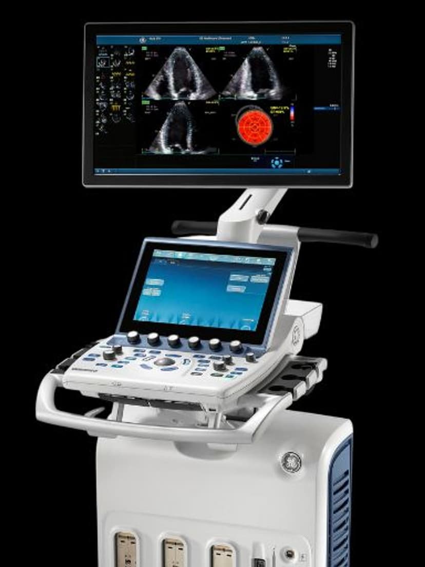
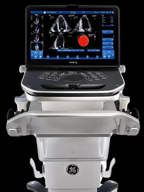
Available on these systems
Our Partners


Want LVO Contrast in your practice?
Request a quote for a system that includes this feature.