
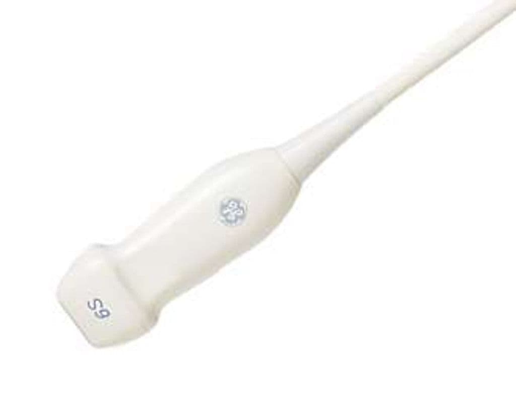
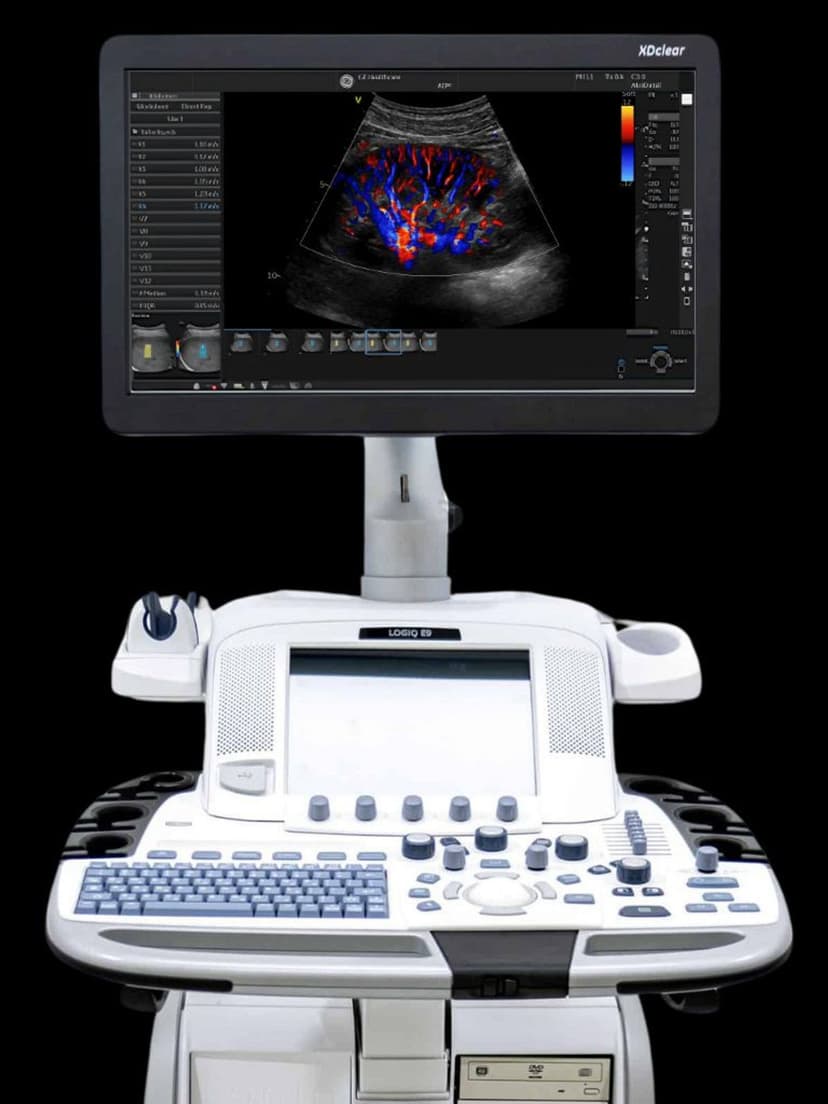
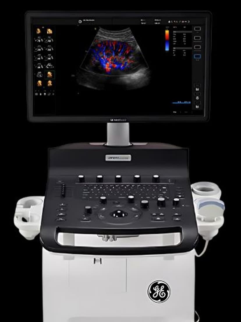
GE 6S-RS
The 6S-RS shifts its frequency range above standard adult cardiac arrays to target the smaller structures in neonatal and pediatric hearts. Its compact footprint fits between neonatal ribs and fontanelles, supporting both echocardiography and cranial imaging through a single probe. RS-pin connectivity pairs the 6S-RS with Vivid, Venue, Versana, and LOGIQ systems for B-Mode, Color Doppler, and PW Doppler across the broadest range of GE platforms.

Specifications
Higher frequency range than adult cardiac phased arrays targets the smaller structures in pediatric and neonatal hearts.
Standard cardiac sector angle captures four-chamber and outflow tract views in pediatric patients.
Sufficient depth for pediatric cardiac and abdominal imaging from neonate through adolescent.
Full Doppler mode set supports hemodynamic quantification in congenital heart disease.
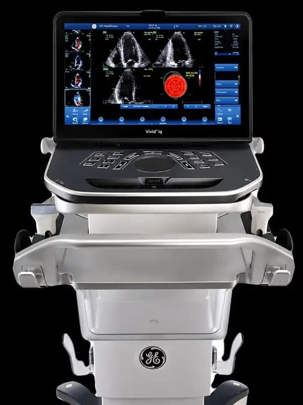
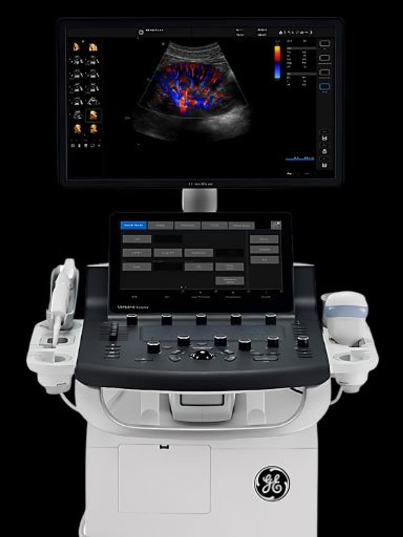
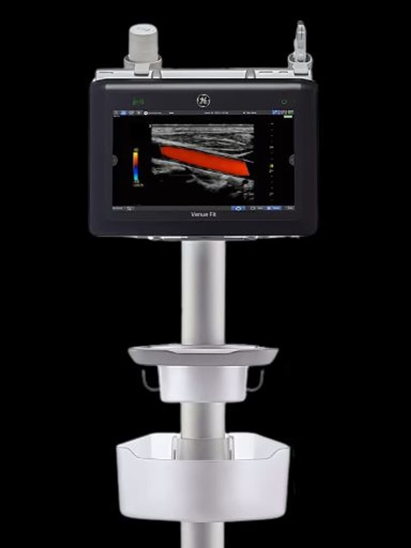
RS-pin interface connects to GE Vivid, Venue, Versana, and LOGIQ RS-compatible platforms.
Applications
Pediatric & Neonatal Echocardiography
The 5–7 MHz upper range resolves intracardiac structures including septal defects, valve leaflets, and coronary origins in neonates and infants. A compact footprint seats between narrow intercostal spaces for parasternal and apical views in patients under 10 kg. PW Doppler quantifies shunt velocities and valve gradients for congenital heart disease workup and post-surgical follow-up.
Neonatal Cranial Imaging
Coronal and sagittal views through the anterior fontanelle detect intraventricular hemorrhage and periventricular leukomalacia in critically ill neonates. The 7 MHz ceiling improves gray-white matter differentiation in the near field. Color Doppler maps the circle of Willis and anterior cerebral artery flow for bedside perfusion assessment in the NICU.
Fetal Cardiac Assessment
Higher operating frequencies compared to standard obstetric probes improve visualization of fetal cardiac chambers, septa, and outflow tracts during targeted fetal echocardiography. Color and PW Doppler detect valve regurgitation and abnormal flow patterns across four-chamber and outflow tract views. The 2.0 MHz low end maintains penetration through maternal tissue at earlier gestational ages.
Our Partners


Need this probe for your system?
Request a quote with pricing, compatibility verification, and delivery timeline.