
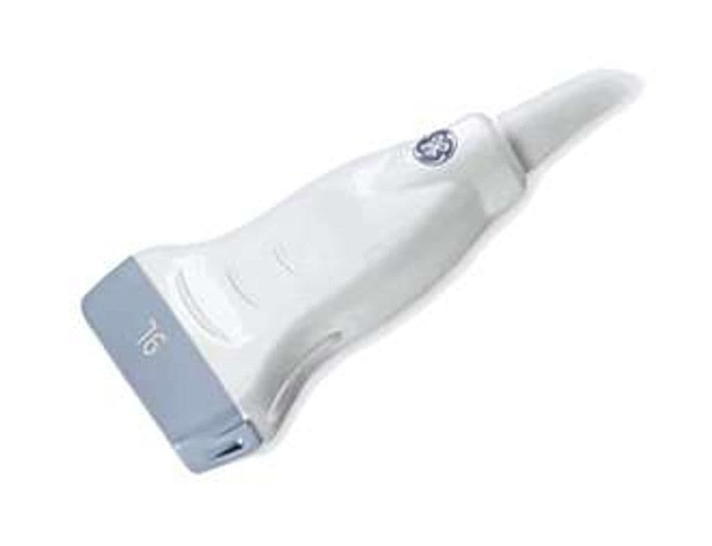
GE 9L-D
The 9L-D pushes linear probe bandwidth lower than most competitors in its class, starting at 2.0 MHz for deeper tissue penetration while still reaching 10 MHz for superficial detail. This makes it a strong choice for practices that need one linear transducer to handle both deeper vascular work and near-field MSK assessment on the same Vivid cardiac platform. Contrast-enhanced ultrasound compatibility on supported systems adds another diagnostic dimension for liver lesion characterization and musculoskeletal perfusion studies.

Specifications
Extended low-frequency range reaches deeper vessels and structures than typical high-frequency linear probes.
Standard diagnostic modes for anatomic and hemodynamic assessment across vascular and MSK applications.
Adequate depth for peripheral vascular work and deeper MSK structures like the hip.
D-Pin interface connects to GE Vivid E95 and Vivid S70 cardiac systems.
Applications
Vascular Imaging
The 2.0 MHz low end reaches deeper vessels that standard high-frequency linears cannot adequately image, including the iliac arteries and deeper segments of the femoral system. Carotid duplex scanning uses the 8–10 MHz range for intima-media thickness measurement and plaque morphology assessment. Spectral Doppler waveform analysis quantifies stenosis severity across the peripheral arterial and venous systems.
Musculoskeletal Assessment
Dynamic evaluation of shoulder, elbow, and ankle pathology benefits from 10 MHz near-field resolution for tendon and ligament visualization. The extended low-frequency range reaches deeper structures like the hip joint and gluteal tendons that are often beyond the range of narrower-bandwidth linear probes. Color Doppler detects neovascularity in tendinopathy and guides therapeutic injections with real-time needle visualization.
Contrast-Enhanced Imaging
On supported Vivid systems, the 9L-D works with contrast-enhanced ultrasound protocols for evaluating focal liver lesions and assessing tissue perfusion in superficial structures. The low-frequency capability at 2.0 MHz supports the low mechanical index needed for microbubble persistence during contrast wash-in and wash-out studies. This adds a diagnostic layer for practices managing patients who need both cardiac and abdominal evaluation.
Our Partners


Need this probe for your system?
Request a quote with pricing, compatibility verification, and delivery timeline.