
GE BE9CS-D
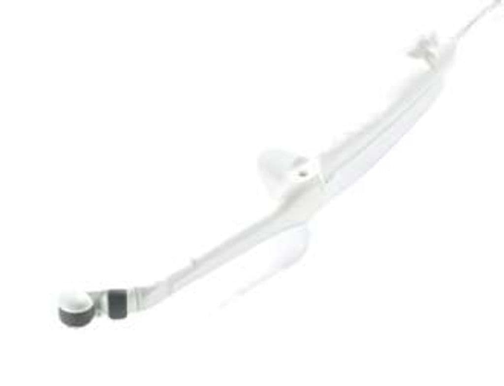
The BE9CS-D is a biplane endocavity transducer that provides two orthogonal imaging planes from a single insertion, eliminating the need to rotate the probe during transrectal prostate or transvaginal pelvic examinations. The 3–12 MHz wideband range delivers high-resolution imaging of the prostate gland, endometrium, ovaries, and pelvic floor musculature. A 127-degree field of view in the sagittal plane captures wide anatomic coverage, and D-Pin connectivity restricts it to GE's flagship LOGIQ platforms where advanced imaging modes are available.
Compatible Systems


Specifications
Wideband endocavity range covers deep prostate imaging through high-resolution endometrial assessment.
Wide sagittal sector captures the full prostate gland or uterus in a single image.
Orthogonal imaging planes from a single insertion reduce exam time and patient discomfort.
Full diagnostic mode set for anatomic and vascular endocavity assessment.
Sufficient depth for transrectal prostate imaging and transvaginal adnexal assessment.
D-Pin interface connects to GE's flagship LOGIQ E10, Totus, and Fortis systems.
Applications
Transvaginal OB/GYN Imaging
High-frequency imaging at 8–12 MHz resolves endometrial thickness, follicular development, and ovarian morphology for fertility assessment and gynecologic pathology screening. The biplane capability displays both sagittal and coronal uterine views without probe rotation, reducing exam time during IVF monitoring. Color Doppler maps adnexal vascularity for characterization of ovarian masses.
Transrectal Prostate Imaging
Biplane imaging captures both sagittal and axial views of the prostate gland for volume estimation and lesion localization. The 127-degree sagittal field of view covers the entire gland from base to apex in a single image. Targeted biopsy guidance benefits from the dual-plane visualization that reduces the need for probe manipulation during sampling.
Pelvic Floor Assessment
The wide frequency range supports both superficial pelvic floor muscle visualization and deeper assessment of the levator ani complex. Dynamic imaging during Valsalva maneuver evaluates pelvic organ prolapse and bladder neck mobility. The microconvex geometry matches the anatomy of the endocavitary approach for consistent acoustic coupling.
Our Partners


Need this probe for your system?
Request a quote with pricing, compatibility verification, and delivery timeline.