
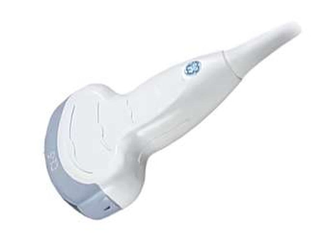
GE C1-5-RS
The C2.0 – 5.0-RS pairs a compact convex footprint with a 2.0 – 5.0 MHz wideband range, giving it enough penetration for deep abdominal organs and enough resolution for second-trimester fetal anatomy. A 60-degree field of view captures broad cross-sections of the liver, kidneys, and gravid uterus in a single sweep. RS-Pin connectivity makes it available across a wide installed base of GE systems including LOGIQ, Venue, Versana, and Vivid portables.

Specifications
Wideband range covers deep abdominal imaging through mid-depth obstetric and pelvic structures.
Wide sector captures broad cross-sections of abdominal organs and the gravid uterus.
Full diagnostic mode set for anatomic and hemodynamic abdominal assessment.
Deep penetration reaches the retroperitoneum and posterior structures in larger patients.
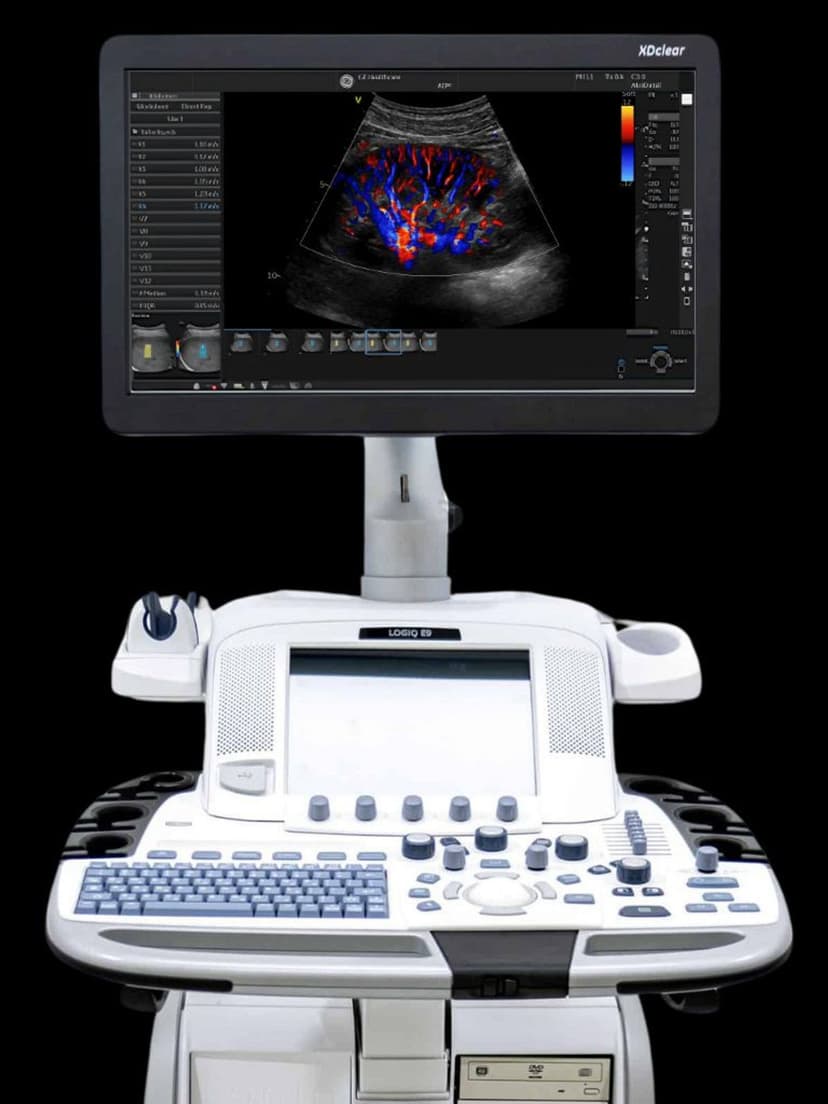
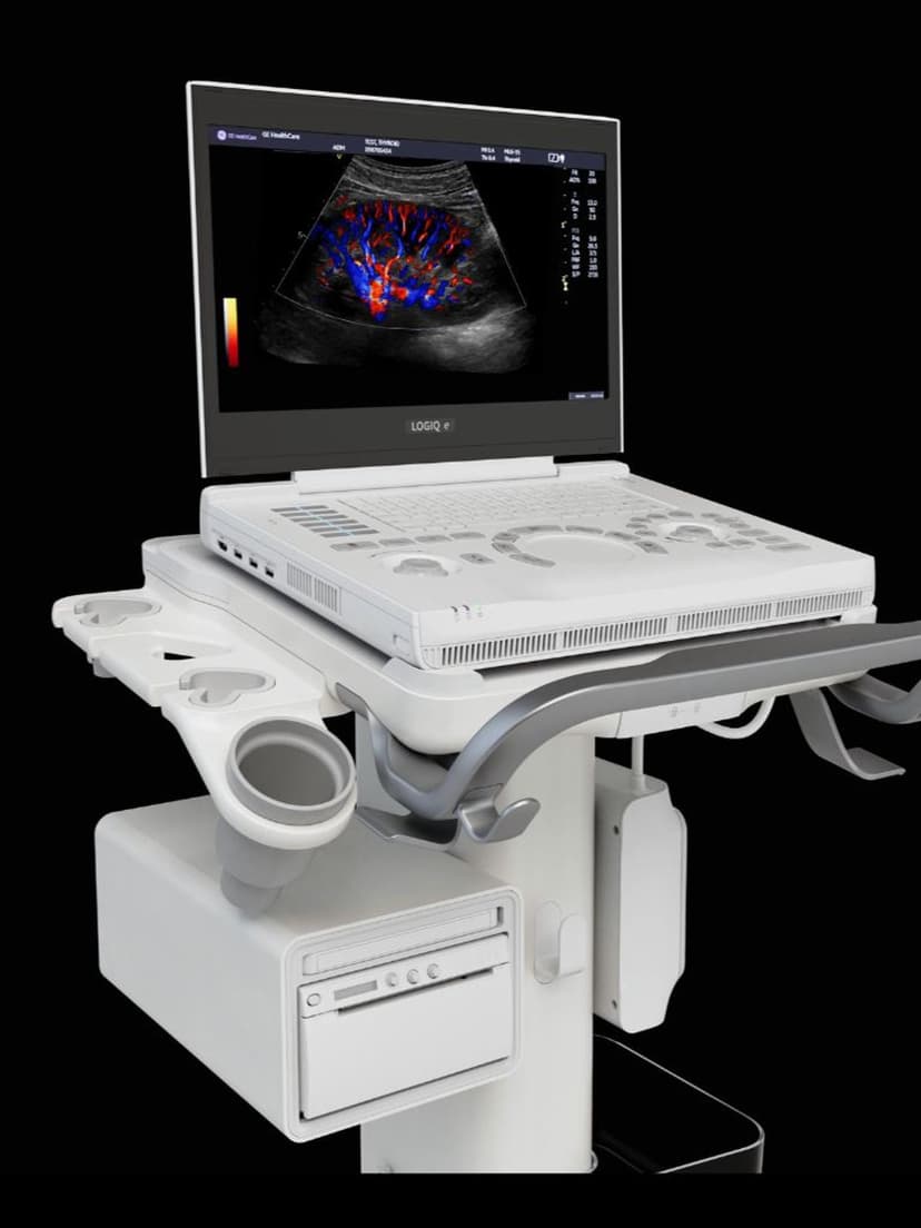
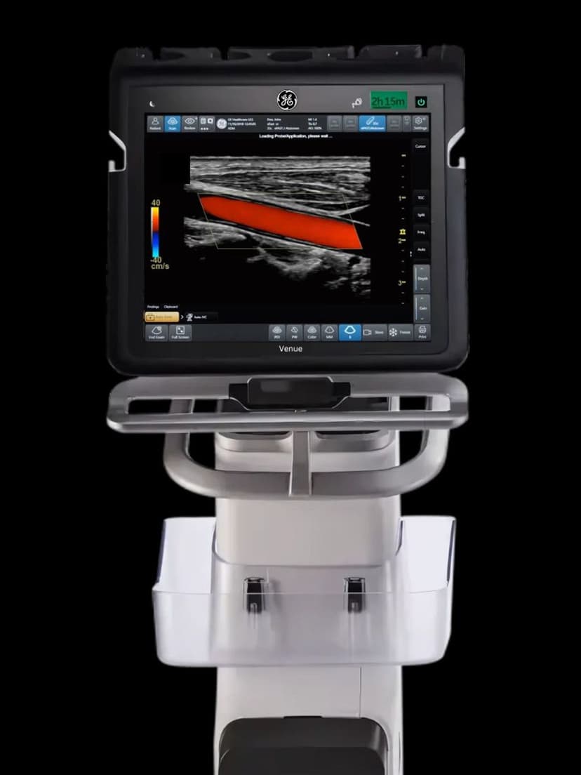
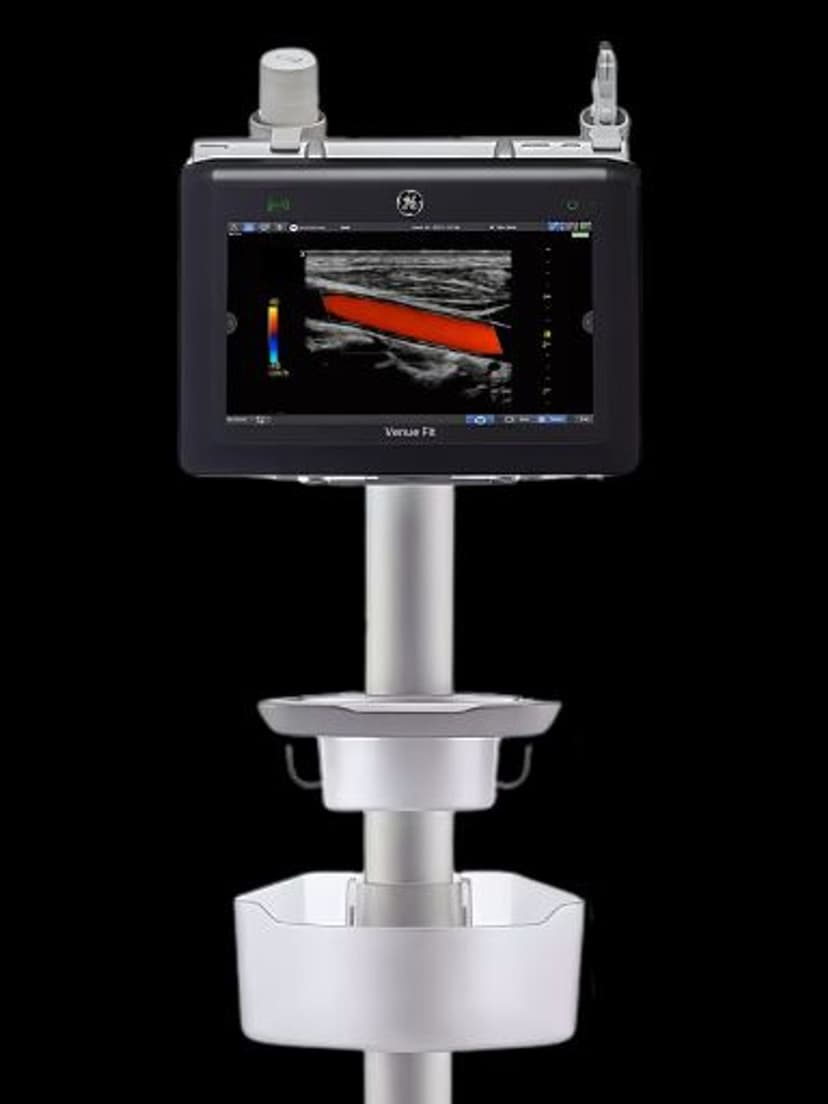
RS-Pin interface connects to GE Venue, Versana, LOGIQ P-series, and Vivid portable systems.
Applications
Abdominal Imaging
The 1.0 MHz low end provides the penetration needed for full-depth liver, renal, and retroperitoneal assessment in larger patients. Color Doppler identifies hepatic and renal vascular flow for portal hypertension screening and transplant follow-up. The 60-degree field of view reduces the number of sweeps needed to survey the right upper quadrant.
OB/GYN Imaging
Frequency range spans standard obstetric imaging from early second trimester through third trimester biometry. The convex geometry matches the curvature of the gravid abdomen for consistent acoustic contact. PW Doppler supports umbilical artery and middle cerebral artery flow velocity measurements used in fetal surveillance protocols.
Urological & Pelvic Imaging
Transabdominal approach to the bladder, prostate, and pelvic structures benefits from the low-frequency penetration. Post-void residual volume measurements and renal calculus detection are supported in B-mode. The compact convex footprint maintains good skin contact in the suprapubic region.
Our Partners


Need this probe for your system?
Request a quote with pricing, compatibility verification, and delivery timeline.