
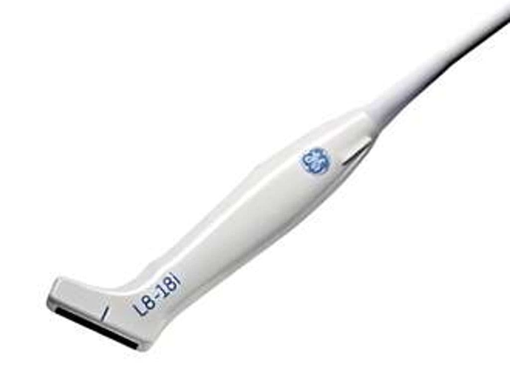
GE L8-18i-D
The L8-18i-D pairs a narrow 25 mm footprint with 5.0 – 18.0 MHz bandwidth for high-resolution imaging of superficial structures in tight anatomical spaces. The compact aperture accesses the neonatal fontanelle, small joints, and intercostal windows where wider probes lose contact. On LOGIQ E10, LOGIQ Fortis, LOGIQ Totus, Vivid E95, and Vivid S70 systems, the L8-18i-D supports B-Mode, Color Doppler, and PW Doppler for both morphology and flow assessment.


Specifications
Extended high-frequency range resolves sub-millimeter structures in thyroid, MSK, and neonatal exams.
Narrow footprint accesses fontanelles, small joints, and tight intercostal spaces.
Sufficient depth for superficial and mid-depth imaging in small parts and pediatric exams.
Full diagnostic mode set for tissue morphology and vascular flow assessment.
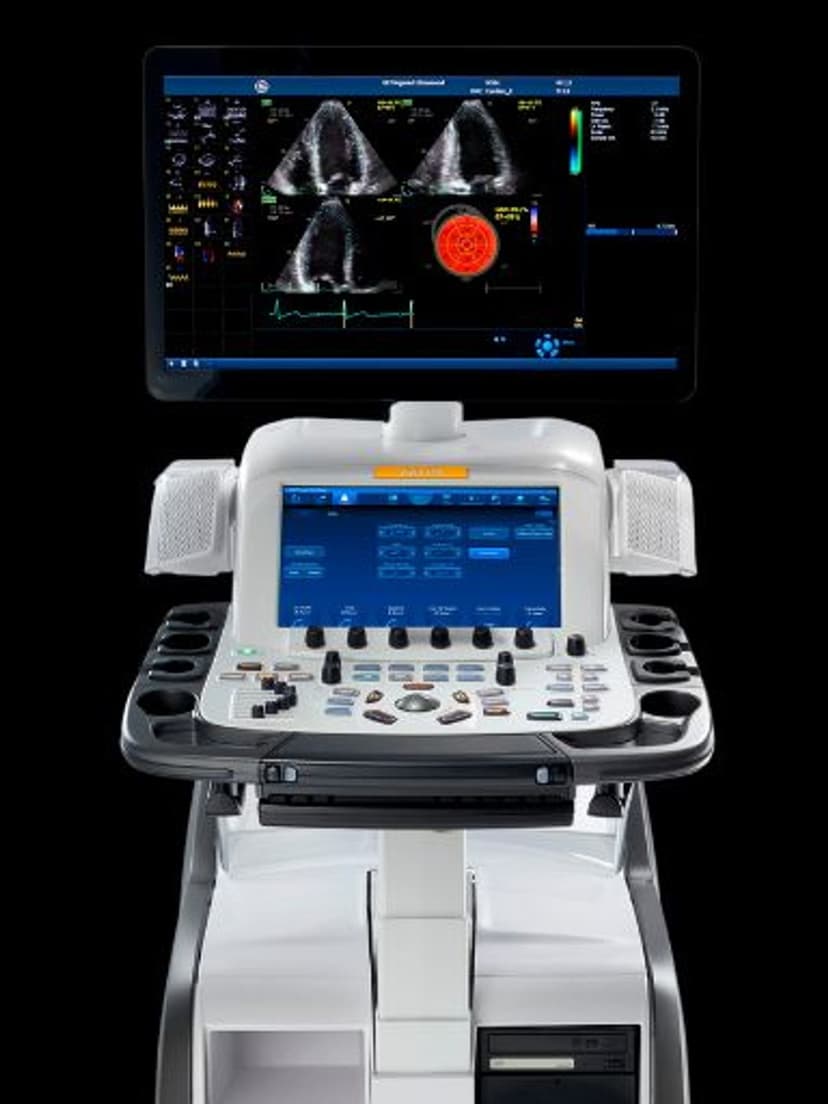
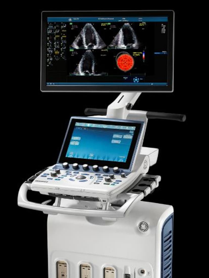
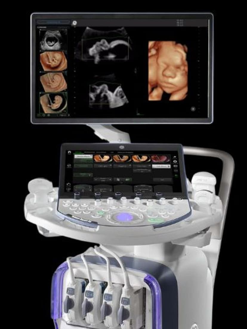
D-Pin interface connects to GE flagship LOGIQ and Vivid systems.
Applications
Superficial Small Parts Imaging
The 15 MHz upper range resolves sub-millimeter structures in the thyroid, salivary glands, and superficial lymph nodes. The 25 mm aperture focuses on individual nodules and lesions without the field-of-view excess of wider linear arrays. Targeted biopsy guidance benefits from the narrow footprint that fits between the needle path and transducer without compromising visualization.
Musculoskeletal Imaging
The compact footprint reaches into tight spaces at the finger, wrist, elbow, and ankle where standard-width linear probes cannot maintain full contact. At 12–18 MHz, tendon fiber patterns, ligament tears, and small joint effusions are visible with sub-millimeter detail. The 5.0 MHz low end provides enough penetration for deeper structures like the median nerve at the carpal tunnel.
Neonatal & Pediatric Imaging
The 25 mm aperture seats on the neonatal anterior fontanelle for cranial imaging without the overhang that larger probes cause on a small head. High-frequency B-mode differentiates gray and white matter for hemorrhage and ischemic injury detection. Pediatric hip, spine, and superficial abdominal imaging benefit from the resolution at 10–18 MHz in patients with minimal overlying tissue.
Our Partners


Need this probe for your system?
Request a quote with pricing, compatibility verification, and delivery timeline.